Electrode Placement (SENIAM)
1/3 from mastoid process to jugular notch
Orientation: Parallel to muscle fiber direction
Source:
Falla et al. 2002 ↗
Falla D, Dall'Alba P, Rainoldi A, Merletti R, Jull G. Location of innervation zones of sternocleidomastoid and scalene muscles - a basis for clinical and research electromyography applications. Clin Neurophysiol. 2002;113(1):57-63.
Methodological background of the source
Falla et al. (2002) mapped the innervation zones of the sternocleidomastoid and scalene muscles using multi-channel surface EMG. The paper explains why the electrodes are deliberately placed away from the innervation zone: over the innervation zone, amplitude cancellation and unstable signals occur. The position is therefore justified electrophysiologically, not just anatomically.
Recording notes per Criswell 2011, site „Sternocleidomastoid (SCM)“, p. 279
- Placement type
- Specific site The signal originates predominantly from this muscle.
- Signal check
- Rotate, side-bend or flex the head.
- Crosstalk from
- upper trapezius · scalenes · cervical neck/head muscle groups
- Artefacts
- ECG pickup, increased if electrodes sit too far anteriorly.
Based on SENIAM recommendations (Hermens et al. 2000). Not an official SENIAM product. See all references →
· How electrodes are placed (guide)
· Glossary
All 48 muscle protocols as a PDF — free
Print-ready for the lab or clinic: 84 pages of protocols (SENIAM + peer-reviewed), plus the SENIAM table, a placement checklist and a sensor comparison sheet as A4 sheets.
Anatomy
Origin
Manubrium of sternum, medial third of clavicle
Insertion
Mastoid process, superior nuchal line
Innervation
Spinal accessory nerve (XI), from the upper five cervical ventral roots
Test Movement
Head rotation to opposite side against resistance
This muscle has no SENIAM-standardized electrode position. Placement should be based on anatomical knowledge.
Measure M. Sternocleidomastoideus activity live
The electrode is placed — what next? easyEMG visualises muscle activity in real time, with SENIAM-compliant placement guidance, up to 4 channels at once and biofeedback training right afterwards.
In therapy, this muscle is usually about tone regulation and consciously relaxing below a threshold.
Frequently Asked Questions
Where do I place the EMG electrode on the M. Sternocleidomastoideus?
1/3 from mastoid process to jugular notch. Orientation: Parallel to muscle fiber direction. Not covered by SENIAM — placement per Falla et al. 2002.
Which test movement activates the M. Sternocleidomastoideus?
Head rotation to opposite side against resistance. It serves as a signal check before recording: with correct electrode placement the EMG amplitude rises clearly during this movement.
Is the M. Sternocleidomastoideus a SENIAM-recommended muscle?
No. The M. Sternocleidomastoideus is not included in the official SENIAM catalogue. The electrode position documented here follows the established protocol per Falla et al. 2002.
Related Muscles in This Region
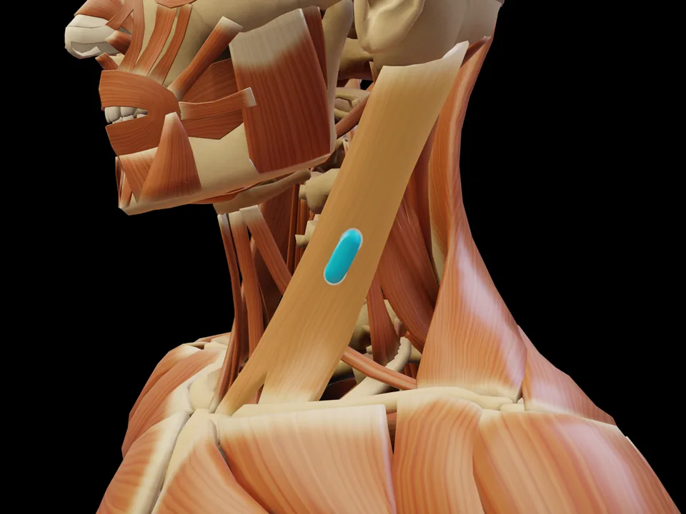 Preview — open the 3D viewer to rotate the model and check neighbouring muscles.
View in 3D Viewer
Preview — open the 3D viewer to rotate the model and check neighbouring muscles.
View in 3D Viewer