Why the Electrode Must Not Sit on the Innervation Zone
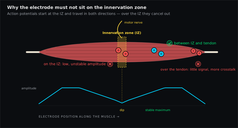
The innervation zone is the band of a muscle where the motor axons end on the fibres and the action potentials are born; from there they travel in both directions towards the tendons. A bipolar surface electrode pair placed over that band sees potentials moving away from it in opposite directions, and their contributions partly cancel: the recorded amplitude is smaller, the spectrum is distorted, and both change sharply when the pair moves by a centimetre. That is why SENIAM places electrodes between the innervation zone and the distal tendon — and why an amplitude comparison made over the innervation zone cannot be trusted.
What happens under the electrodes
A single muscle fibre is excited at its neuromuscular junction. The depolarisation then propagates along the fibre at 3–5 m/s (see conduction velocity) in both directions until it reaches the tendon and extinguishes. A bipolar pair aligned with the fibre records the difference between the two electrodes; a potential travelling along the fibre passes electrode 1, then electrode 2, and produces a clean biphasic waveform.
Now put the pair over the innervation zone. The potential starts between the electrodes and runs outward — towards electrode 1 on one side and electrode 2 on the other, at the same time. The two electrodes see nearly the same thing at nearly the same time, and a differential amplifier subtracts what both share. The result is what Cram's (following Fridlund & Cacioppo) describes as "lower amplitudes owing to differential amplification", and what the amplitude cancellation entry in the glossary summarises: signal that is physiologically there but cancels at the electrodes.
Three practical consequences follow:
- The amplitude is smaller than the muscle's activity warrants — and by an amount that depends on exactly where the innervation zone lies relative to the two electrodes.
- It is unstable. The innervation zone shifts under the skin when the muscle shortens or lengthens (Konrad's electrode-migration problem in reverse); a joint angle change can move the pair on or off the zone and change the amplitude by tens of percent without any change in activation.
- The frequency content is distorted, so median-frequency fatigue analysis over the innervation zone is unreliable too.
Innervation zones are not points but bands, often several centimetres wide, and their location varies between people. Some muscles have one, some several (the trapezius, the biceps with its two heads).
How SENIAM avoids it
SENIAM's sensor-placement procedure defines each site as a fraction of a line between two palpable landmarks — chosen, muscle by muscle, so that the pair lands on the belly between the innervation zone and the distal tendon, on fibres that run in a well-defined direction. Two examples from the EMG Guide muscle pages:
- Biceps Brachii: on the line from the medial acromion to the cubital fossa, at one third from the fossa — distal to the mid-belly, where the main innervation zone lies.
- Tibialis Anterior: at one third on the line from the fibular head to the medial malleolus — proximal third, but off the innervation zone that lies more proximally.
Rainoldi and colleagues (2004) mapped the innervation zones of thirteen lower-limb muscles with linear electrode arrays and gave, for each, an optimal placement window — independent confirmation that the SENIAM fractions for Vastus Medialis, Vastus Lateralis or Gastrocnemius (Medial Head) sit clear of the zones. Falla et al. (2002) did the same for the Sternocleidomastoid and the scalenes and recommended positions that avoid the zones — the source of the EMG Guide placement for both. Barbero, Merletti and Rainoldi's atlas (2012) collects such maps for the whole body.
The lesson is uniform: the standard positions look arbitrary ("one third", "50 %") only until you know that each fraction is where the innervation zone is not.
Muscles that are notorious
- Upper trapezius. SENIAM places the Upper Trapezius pair at 50 % between C7 and the acromion; Cram's, citing Veiersted, prefers a slight lateral shift from that midpoint for a stronger, more reliable signal. The two are close — what matters is not to drift medially, where amplitude becomes unstable, and to keep the same position every session.
- Biceps brachii. Innervation zone around mid-belly; a "central" placement is exactly wrong. Distal third.
- Sternocleidomastoid. Falla 2002: innervation zones located along the muscle; the muscle also changes length by 50 % during rotation (Cram's), so a placement that is clear of the zone in neutral may not be in rotation.
- Vastus medialis, vastus lateralis, gastrocnemius. Multiple or oblique zones; Rainoldi 2004 gives the recommended windows.
- Brachioradialis. EMG Guide's position follows the scoping review by Merlo et al. (2021), which pools the evidence on electrode size and placement for this muscle — including where its innervation zone lies — and recommends the proximal belly.
How to spot the problem in your own recording
- Palpate and mark the SENIAM fraction first; do not "centre" the pair on the belly by eye.
- Test contraction: the amplitude should be large and repeatable. If it is small for the effort and rises noticeably when you slide the pair 1–2 cm along the fibres, you were on the zone. Move distally.
- Change the joint angle slightly and repeat. A stable placement changes little; a placement on or next to the zone changes a lot.
- Look at the raw waveform. Over the belly you see the typical broad biphasic bursts; over the zone the trace is smaller with sharper, higher-frequency content.
- If you have a linear array or multiple channels, the innervation zone is where the propagating potentials reverse polarity — you can see it directly.
Muscles referenced in this article
From placement to measurement
EMG Guide shows where the electrodes go. easyEMG with PicoBlue sensors visualises the signal live afterwards — SENIAM-conform, in real time, with a signal check on screen.
Frequently asked questions
Is the innervation zone the same as the motor point?
Closely related. The motor point is the skin location where the muscle is most easily excited by electrical stimulation — the point of lowest stimulation threshold, usually over the innervation zone. EMS/NMES pads target it; EMG detection electrodes avoid it. Same anatomy, opposite intent (see the placement guide FAQ).
How far from the innervation zone should the pair be?
Far enough that both electrodes lie on the same side of it, on fibres travelling in one direction — in practice the SENIAM landmark fraction, or the window given by Rainoldi 2004 for lower-limb muscles. There is no fixed centimetre value; the zone's width and position vary between people.
Does this matter for biofeedback, or only for research?
It matters whenever amplitude is compared — between sides, sessions or before/after treatment. Biofeedback trend displays are exactly such comparisons. It matters less for on/off timing.
Can a filter or normalisation fix a placement on the innervation zone?
No. Normalisation cancels detection-condition effects only if the reference and the test share the same effect; a placement that shifts on and off the zone with joint angle does not. Move the electrodes.
Sources
- Hermens HJ et al. Development of recommendations for SEMG sensors and sensor placement procedures. J Electromyogr Kinesiol. 2000;10:361–374.
- Rainoldi A, Melchiorri G, Caruso I. A method for positioning electrodes during surface EMG recordings in lower limb muscles. J Neurosci Methods. 2004;134:37–43.
- Falla D, Dall'Alba P, Rainoldi A, Merletti R, Jull G. Location of innervation zones of sternocleidomastoid and scalene muscles — a basis for clinical and research electrode placement. Clin Neurophysiol. 2002;113:57–63.
- Barbero M, Merletti R, Rainoldi A. Atlas of Muscle Innervation Zones. Springer; 2012.
- Criswell E. Cram's Introduction to Surface Electromyography. 2nd ed. 2011 — Ch. 4 (p. 70), Ch. 5 (p. 75).
- Konrad P. The ABC of EMG. Noraxon; 2005 — p. 17–18.
- Merlo A, Bò MC, Campanini I. Electrode size and placement for surface EMG bipolar detection from the brachioradialis muscle: a scoping review. Sensors. 2021;21:7322.
Read next
EMG Electrode Placement — The Practical Guide
How to place surface EMG electrodes correctly — SENIAM rules, skin prep, 20 mm spacing, fibre direction, reference electrode, signal check and common mistakes.
10 Factors That Distort Your EMG Signal
Skin, fat, temperature, spacing, innervation zone, crosstalk, ECG, motion, cables, fatigue — ten influences that change an EMG amplitude, and how to control each.
Surface EMG Glossary
54 terms of surface electromyography explained in one sentence each — from amplitude cancellation to volume conduction. With sources (SENIAM, Cram's, Konrad).
All articles →