10 Factors That Distort Your EMG Signal
A surface EMG amplitude is not a property of the muscle alone. Between the muscle fibres and the number on the screen lie skin, fat, electrode geometry, cables, the amplifier and the environment — and each of them can change the value without any change in muscle activation. Konrad summarises it as the "detection condition"; Cram's devotes a chapter to the factors that affect interpretation. Here are the ten that matter most in practice, what each does to the signal, and how to keep it under control.
1. Skin condition and preparation
Dead skin cells, sweat, oil and hair raise and destabilise the skin impedance. High or unequal impedance under the two electrodes reduces the amplifier's ability to cancel interference — the baseline gets noisy, mains hum and motion artifacts get through, and small activations disappear in the noise. Cram's: poor electrode contact artificially lowers SEMG amplitudes.
Control: shave, abrade lightly, clean with alcohol, wait a few minutes before recording (impedance keeps falling). Slightly reddened skin is the visual target. Detail in the placement guide.
2. Subcutaneous fat
Adipose tissue insulates and attenuates. Cram's reports a correlation of about 0.5 between skinfold thickness and resting SEMG level, and about 0.25 during moderate contraction — fat affects quiet, low-amplitude recordings more than active ones. Fat distribution differs between people, between sexes and between sites on the same body, so absolute microvolt values are not comparable across any of those.
Control: you cannot remove it, only account for it. Note skinfold thickness at the site, compare within the person (left vs. right, before vs. after) rather than across people, and normalise (see factor 10).
3. Temperature
Muscle temperature changes conduction velocity: a warm muscle conducts faster, which shifts the power spectrum to higher frequencies and slightly changes amplitude. Cold skin also raises impedance.
Control: let the person acclimatise to the room, do a standardised warm-up before baseline measurements, keep conditions similar between sessions and note room temperature.
4. Electrode size and inter-electrode distance
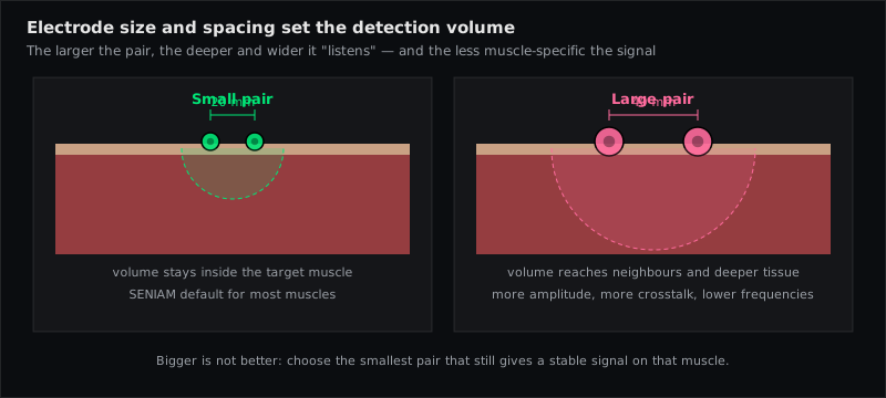
Cram's rule: depth and area of the pickup are proportional to the inter-electrode distance. A wider spacing gives a larger amplitude — and more of the neighbouring muscles. Larger electrodes average more tissue and lower selectivity. Both change the value without changing the muscle.
Control: Ag/AgCl electrodes ≤ 10 mm, 20 mm spacing (SENIAM), identical on both sides and in every session. Write it down.
5. Position relative to the innervation zone and tendon
Over the innervation zone action potentials travel in opposite directions and partly cancel; the amplitude is smaller and unstable and shifts with tiny repositioning. Towards the tendon the fibres thin out and the signal fades. Cram's (after Fridlund & Cacioppo): avoid straddling the motor end-plate region; slightly off-centre is better.
Control: use the SENIAM landmark fraction, which is chosen to sit between innervation zone and distal tendon; verify with a test contraction.
6. Muscle length, joint position and electrode migration
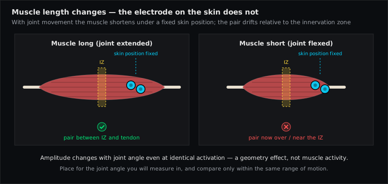
The muscle moves under the skin; the electrodes stay with the skin. Cram's describes the sternocleidomastoid changing its resting length by 50 % during head rotation, so the pair drifts ahead of or behind the belly. Konrad names the Biceps Brachii and Vastus Medialis as typical migrators, and skin stretch as a problem for Rectus Abdominis, the erector spinae and the Upper Trapezius. Even without migration, the same activation produces different amplitudes at different muscle lengths.
Control: place electrodes with the muscle in the position it will mostly be in (Konrad: the most shortened position for dynamic tasks); compare amplitudes only at the same joint angle; prefer isometric or same-range comparisons; document position and posture.
7. Crosstalk from neighbouring muscles
Crosstalk is real EMG from the wrong muscle. It cannot be seen in the raw trace and cannot be filtered out afterwards. Cram's warns that a single pair over one muscle can lead to an erroneous conclusion about that muscle, and grades every atlas site by how prone it is (see placement types).
Control: small electrodes, 20 mm spacing, placement over the belly away from the border, functional test against the neighbours' actions, and — Cram's advice — record synergists or antagonists on further channels so that the pattern exposes it.
8. Physiological artifacts: ECG and breathing
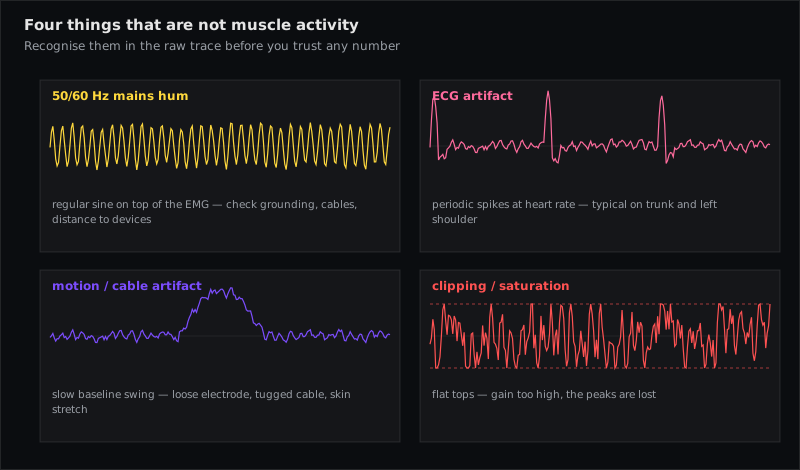
The heart is a large muscle with a large signal. On the trunk and shoulders — Longissimus Thoracis, Rectus Abdominis, Pectoralis Major (Sternocostal Head), the left Upper Trapezius — the ECG appears as regular spikes about once per second and inflates every amplitude measure. Breathing moves the chest wall and shifts the baseline of trunk channels.
Control: recognise the rhythm in the raw signal; place trunk electrodes as far from the heart as the protocol allows; use ECG-reduction algorithms or gating where available; report that the channel is affected.
9. Motion artifacts, cables and mains hum
Relative movement between electrode, cable and skin produces low-frequency baseline waves and sharp spikes; unfixed cables act as antennas for 50/60-Hz hum; poorly grounded devices nearby (treadmills, training machines) inject noise. Konrad's baseline criteria: random spikes below 10–15 µV, mean rectified noise around 1–3.5 µV, bursts returning to zero within milliseconds — anything else is an artifact, not muscle.
Control: thorough skin preparation, cables taped or netted without tension, reference electrode on bone, devices grounded, a 20-Hz high-pass in dynamic tasks, and a look at the power spectrum for a peak at 50/60 Hz before you reach for a notch filter.
10. Fatigue, learning and the day itself
A fatiguing muscle shows a rising amplitude at constant force and a falling median frequency; a person who has learned the task recruits differently than in the first trial; hydration, time of day and previous exercise all change the numbers. None of this is an error — but all of it masquerades as a "difference" if it is not controlled.
Control: standardise order, rest periods and instructions (Konrad's test-standardisation list); randomise or counterbalance conditions; use normalisation to a reference contraction recorded in the same session, so that changes in the detection condition cancel out.
The one principle behind all ten
Absolute microvolts describe the detection condition as much as the muscle. Every comparison — between sides, between sessions, between people — is only as good as the match between the two detection conditions. Keep everything you can identical, document what you cannot, and normalise. Cram's puts it bluntly: SEMG is not a measure of force, strength or effort; it is a measure of the electrical activity that reaches the electrodes.
Muscles referenced in this article
From placement to measurement
EMG Guide shows where the electrodes go. easyEMG with PicoBlue sensors visualises the signal live afterwards — SENIAM-conform, in real time, with a signal check on screen.
Frequently asked questions
Which factor causes the biggest errors in practice?
In our experience the innervation zone and crosstalk — because both produce clean-looking traces that are simply wrong for the muscle you think you are recording. Skin preparation and cables cause the visible problems; these two cause the invisible ones.
Can I compare µV values between two people?
No. Fat, skin, muscle geometry, electrode position and equipment differ; only normalised values (e.g. % of a reference contraction) or patterns and timing are comparable across people.
Does a notch filter solve mains hum?
It removes the 50/60-Hz component — including the real EMG at those frequencies. Grounding, cable fixation and the reference electrode solve the cause; the notch filter should be the last resort, and its use must be reported.
How do I know whether a difference is real?
Repeat the measurement under identical conditions and see whether it persists; check the raw trace for artifacts; make sure the joint angle and task were the same; and ask whether a neighbouring muscle could explain it. Only then interpret.
Sources
- Criswell E. Cram's Introduction to Surface Electromyography. 2nd ed. 2011 — Ch. 5 "Factors That Affect Interpretation" (pp. 75–80), Ch. 3 "Noise and Artifact" (pp. 54–57), Ch. 4 "Site Preparation" (p. 69).
- Konrad P. The ABC of EMG. Noraxon; 2005 — pp. 11 (detection conditions), 14 (skin preparation), 18 (electrode migration), 21–24 (signal check, artifacts).
- Hermens HJ et al. SENIAM recommendations. J Electromyogr Kinesiol. 2000;10:361–374.
- De Luca CJ. The use of surface electromyography in biomechanics. J Appl Biomech. 1997;13:135–163.
Read next
EMG Electrode Placement — The Practical Guide
How to place surface EMG electrodes correctly — SENIAM rules, skin prep, 20 mm spacing, fibre direction, reference electrode, signal check and common mistakes.
Specific, Quasi-Specific, General — The Three Types of Surface EMG Placement
Why there is no single correct electrode position — Cram's three placement types, what each records, where SENIAM fits, and what it means for interpretation.
Surface EMG Glossary
54 terms of surface electromyography explained in one sentence each — from amplitude cancellation to volume conduction. With sources (SENIAM, Cram's, Konrad).
All articles →