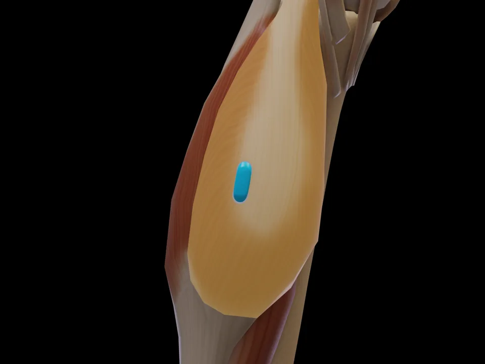
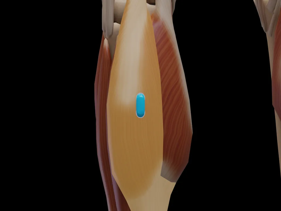
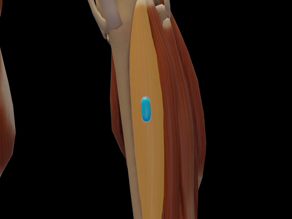
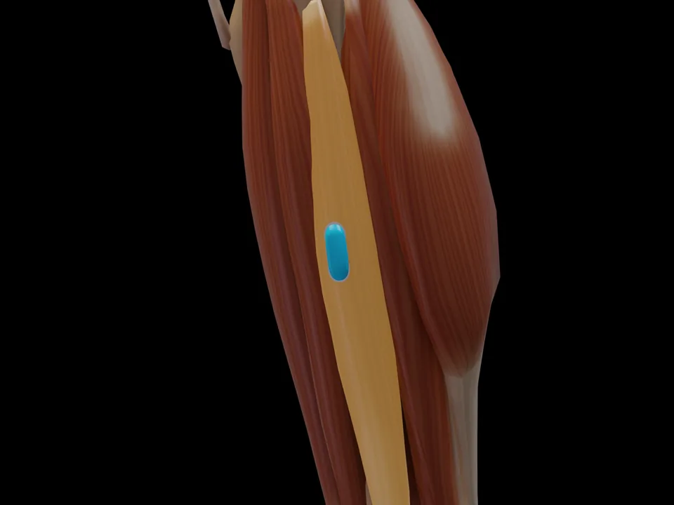
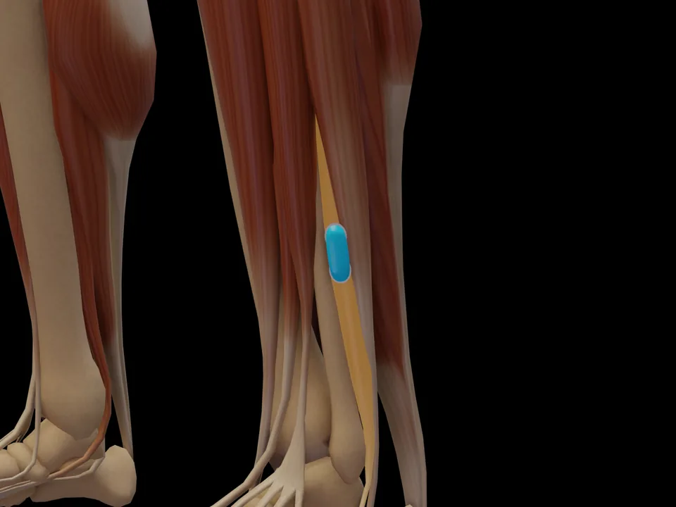
Recording notes & source
per Criswell 2011, site „Soleus“, p. 374
- Placement type
- Quasi-specific site Neighbouring muscles contribute noticeably to the signal — allow for crosstalk.
- Signal check
- Lean forward while standing.
- Crosstalk from
- gastrocnemius
Source:
SENIAM (Hermens 2000) ↗
Hermens HJ, Freriks B, Disselhorst-Klug C, Rau G. Development of recommendations for SEMG sensors and sensor placement procedures. J Electromyogr Kinesiol. 2000;10(5):361-74.
Based on SENIAM recommendations (Hermens et al. 2000). Not an official SENIAM product. See all references →
· How electrodes are placed (guide)
· Glossary
All 49 muscle protocols as a PDF — free
Print-ready for the lab or clinic: 79 pages of protocols (SENIAM + peer-reviewed), plus the SENIAM table, a placement checklist and a sensor comparison sheet as A4 sheets.
Anatomy & Function
The M. Soleus (Soleus) is a muscle of the lower leg region. It is responsible for ankle plantar flexion (single-joint, knee-independent), postural stabilization in standing. This muscle is one of 26 with official SENIAM-recommended surface EMG electrode placement. This page documents the exact electrode position, anatomical attachments and a standardized test movement based on Hermens et al. (2000).
Function
Ankle plantar flexion (single-joint, knee-independent), postural stabilization in standing
Origin
Fibular head, proximal third of the fibula (dorsal), soleal line of the tibia
Insertion
Calcaneus (via Achilles tendon / tendo calcaneus, together with gastrocnemius)
Innervation
Tibial nerve, L5–S2
Clinical applications (SENIAM)
- Gait analysis and foot drop assessment
- Achilles tendinopathy monitoring
- Proprioceptive training after ankle injury
Test Movement
Plantar flexion against resistance with flexed knee (isolated from gastrocnemius)
MVC reference test (Konrad 2005)
- Position
- Seated, knee flexed to about 90°, unilateral plantar flexion.
- Fixation
- Very rigid fixation of the knee (load or machine pad on the thigh) because of the high forces.
- Resistance
- Fixed footplate or load on the knee, plantar flexion against it.
- Control exercise
- Important check exercise for the gastrocnemius, which is at a disadvantage with the knee flexed.
With the knee flexed the gastrocnemius is shortened, so the soleus dominates the plantar flexion.
Konrad P. The ABC of EMG. Noraxon; 2005, p. 32. Konrad describes this test at group level; it applies to all muscles of the group. · Why and how to normalise to MVC
Measure M. Soleus activity live
The electrode is placed — what next? easyEMG visualises muscle activity in real time, with SENIAM-compliant placement guidance, up to 4 channels at once and biofeedback training right afterwards.
In screening, this muscle is usually recorded together with its synergists — running several channels in parallel saves the most time.
Frequently Asked Questions
Where do I place the EMG electrode on the M. Soleus?
2/3 on line from medial femoral condyle to medial malleolus. Orientation: Parallel to the longitudinal axis of the lower leg. Based on the official SENIAM recommendations (Hermens et al. 2000).
Which test movement activates the M. Soleus?
Plantar flexion against resistance with flexed knee (isolated from gastrocnemius). This movement is part of the SENIAM protocol for standardized contraction verification.
Is the M. Soleus a SENIAM-recommended muscle?
Yes. The M. Soleus is one of 26 muscles for which the SENIAM project has published official surface EMG electrode placement recommendations.
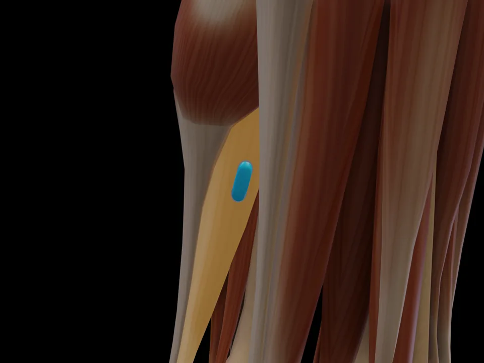 Preview — open the 3D viewer to rotate the model and check neighbouring muscles.
View in 3D Viewer
Preview — open the 3D viewer to rotate the model and check neighbouring muscles.
View in 3D Viewer