MVC Normalisation — Why Microvolts Alone Say Nothing
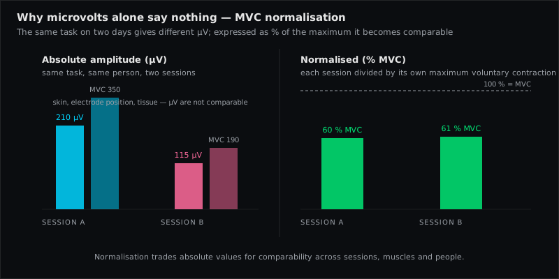
Normalisation expresses a surface EMG amplitude as a percentage of a reference contraction — most often the maximum voluntary (isometric) contraction, MVC or MVIC — instead of in absolute microvolts. It is necessary because the microvolt value depends on the detection condition (skin, fat, electrode position, equipment) as much as on the muscle: the same activation gives different microvolts on different days, sides, muscles and people. Normalisation removes that dependency and rescales the amplitude to "percent of maximum innervation capacity" (Konrad). It changes the y-axis, never the shape of the curve.
The problem normalisation solves
Konrad calls the microvolt scale "uncertain": it varies between electrode sites, between subjects and even between day-to-day recordings of the same site. Cram's spells out the consequence: you should not compare the raw amplitude of the upper trapezius with that of the lower trapezius — a difference may reflect muscle mass under the electrodes rather than effort. To compare across muscles, sessions or people you must normalise first.
The ten factors that distort the signal are the list of things a reference contraction absorbs: if the reference is recorded with the same electrodes, in the same session, under the same skin condition, then dividing by it cancels all of them.
The concept
- For each muscle, record a reference contraction under standardised conditions — usually a maximal isometric effort against fixed resistance.
- Take the amplitude of that reference as 100 %.
- Express every value in the test trials as a percentage of it.
The reference amplitude is not the single highest sample (too variable). Konrad: use the mean of the highest signal portion of e.g. 500 ms, found with a moving window. RMS or a smoothed envelope, same processing for reference and test.
Running an MVC test that is worth the name
Konrad's practical rules (pp. 30–31):
- One test per muscle, in a position that produces maximal innervation — not maximal force output. For limb muscles: isolated single-joint action, statically fixed at mid-range. For trunk muscles: an exercise that activates the whole muscle chain.
- Rigid fixation. Belts, benches, training machines. Without fixation the effort leaks into other segments and the target never reaches its maximum.
- Warm up 5–10 minutes (stretching, light aerobic work).
- Ramp, hold, release: increase force slowly over 3–5 s, hold the maximum for 3 s, release over 3 s. Repeat at least once with 30–60 s rest. Take the higher value.
- Try several candidates for trunk and hip muscles. Konrad's example: for rectus abdominis, external oblique and rectus femoris, different subjects reached their maximum in different exercises — run two or three, in random order, and take the highest.
- Verbal encouragement and feedback. Untrained people rarely produce a true maximum at the first attempt.
- Record everything in one file, pause between positions, and let the software find the peak portions automatically.
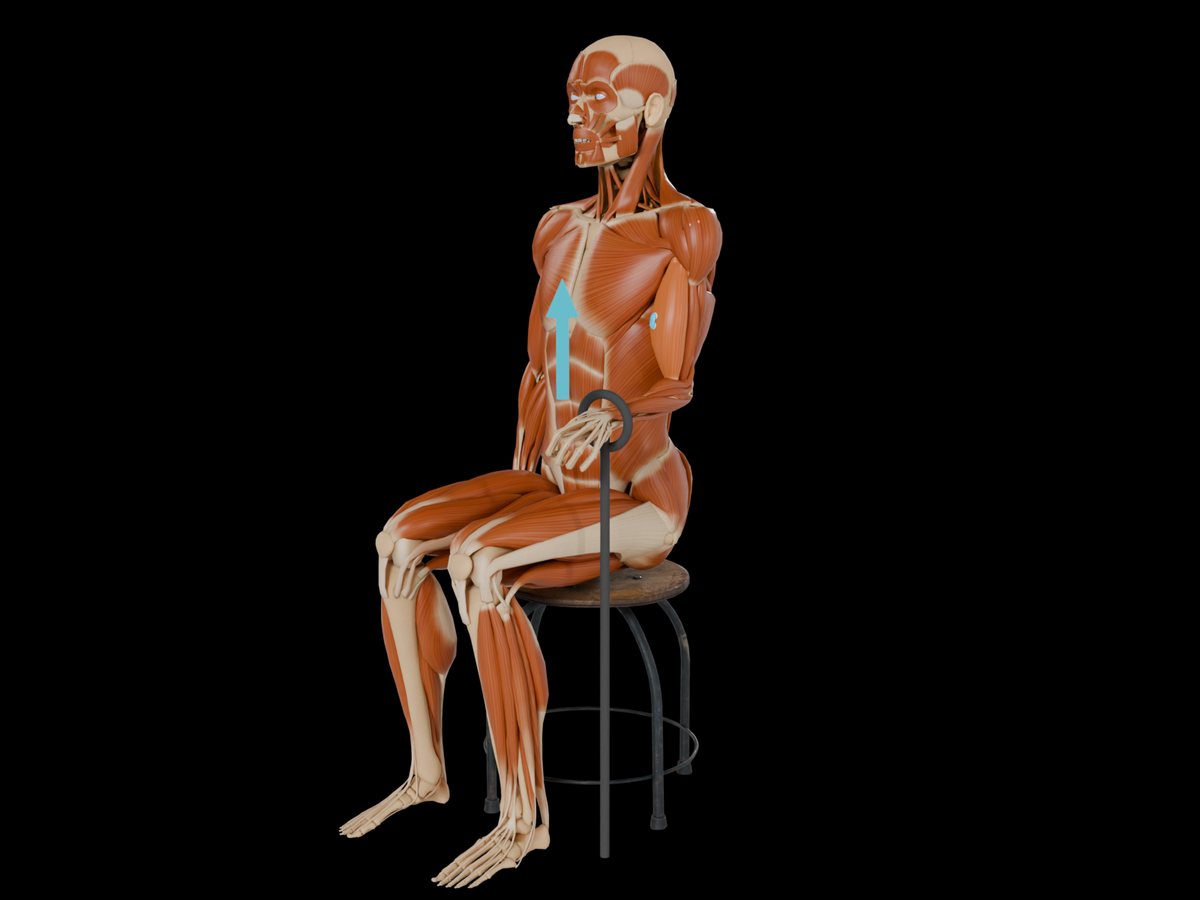
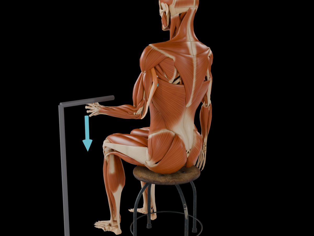
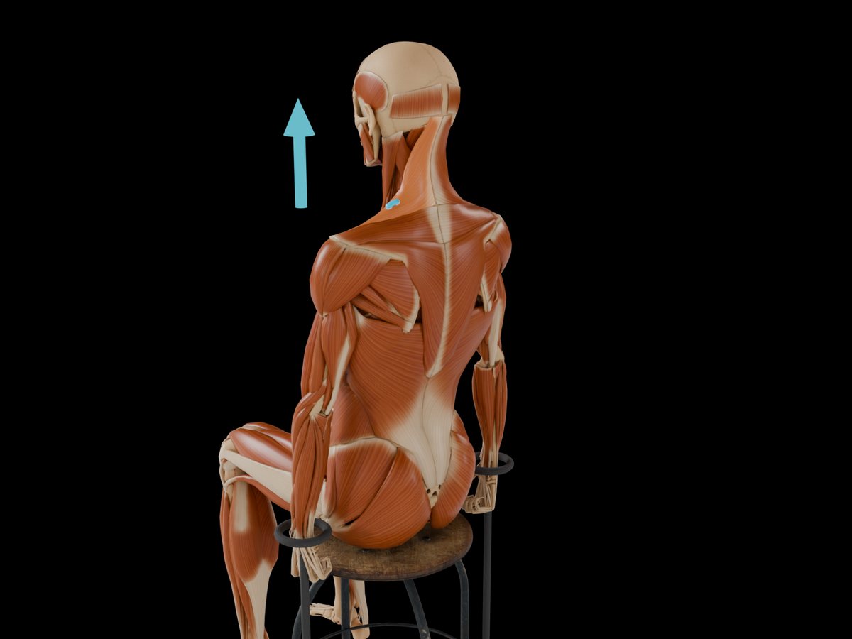
Typical positions from Konrad's table: Biceps Brachii — seated or kneeling, elbow ~90°, forearm and trunk fixed, resistance under the wrist; Triceps Brachii (Long Head) — same set-up, resistance from above; Middle Deltoid — seated, back supported, arms abducted to 90° against fixed straps, bilaterally; Upper Trapezius — shoulder elevation against a fixed arm or a heavy load; Pectoralis Major (Sternocostal Head) — press against a fixed bar in prone or a push-up position, elbow 90°; Rectus Femoris — seated knee extension at ~60° flexion against a fixed strap; Gastrocnemius (Medial Head) — plantar flexion against a fixed footplate, knee extended.
 Biceps Brachii
 Triceps Brachii (Long Head)
 Middle Deltoid
 Upper Trapezius
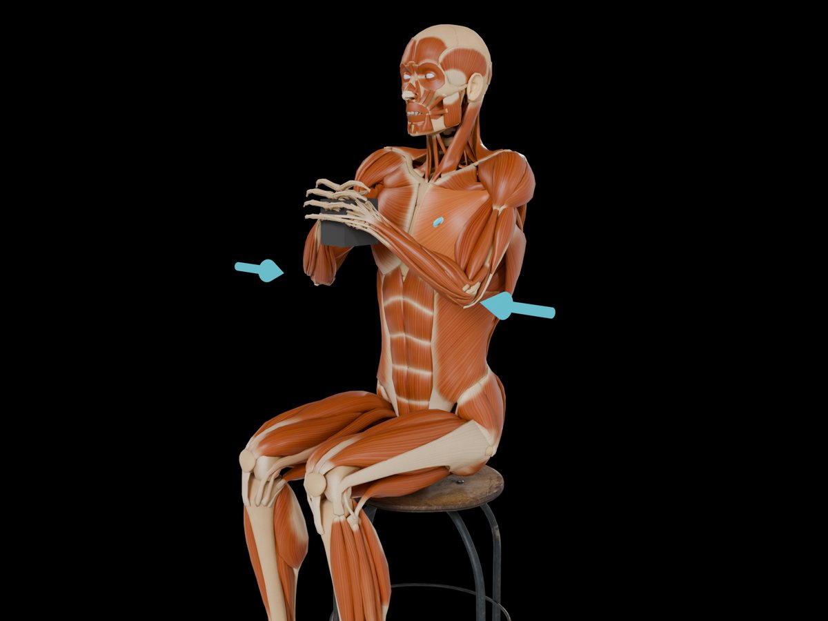 Pectoralis Major (Sternocostal Head)
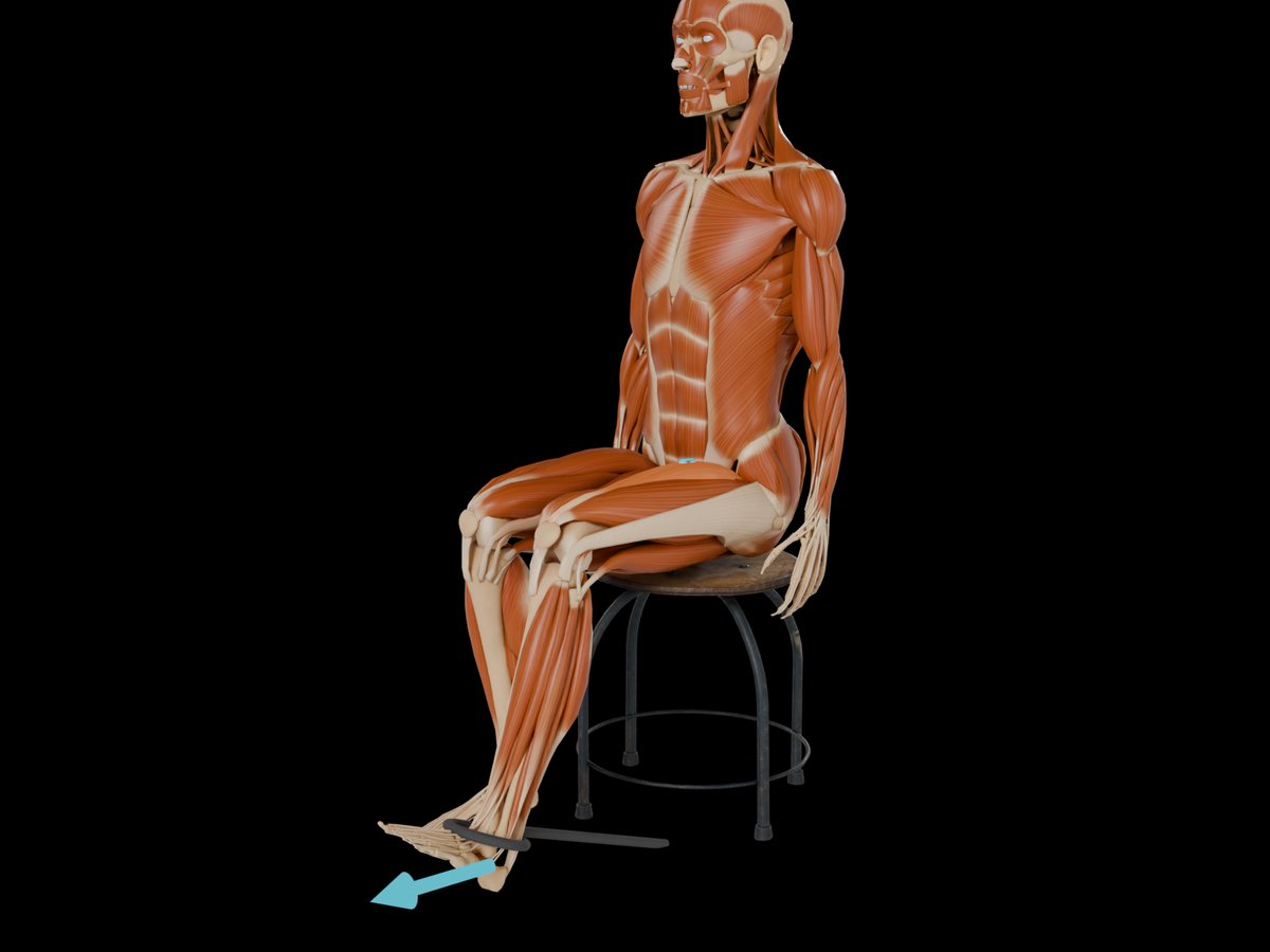 Rectus Femoris
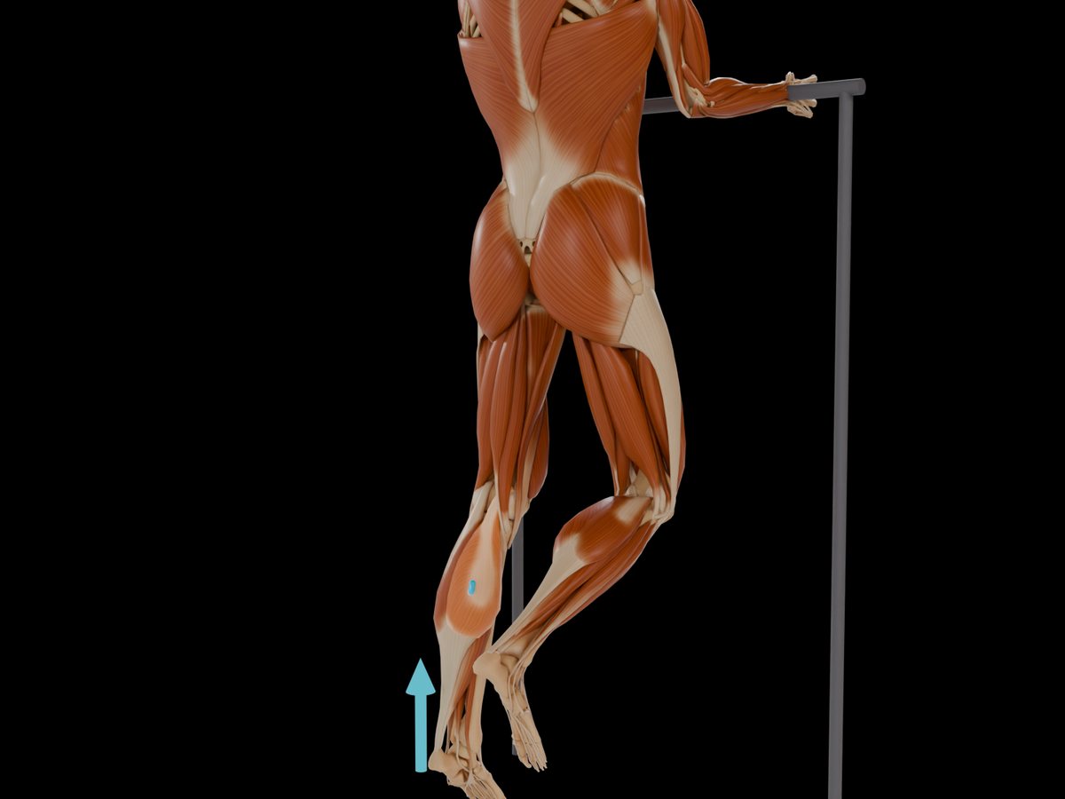 Gastrocnemius (Medial Head)
 Rectus Abdominis
When MVC does not work — and what to do instead
Patients. Konrad is unambiguous: patients cannot and should not perform maximal efforts with injured structures. Pain inhibition also means that even a willing patient's "maximum" is not the physiological maximum, and it changes from day to day. Konrad's clinical alternative is the acceptable maximum effort (AME) — a guideline value for biofeedback, explicitly not an MVC replacement and not comparable across sessions.
Submaximal reference (RVC). Normalise to the EMG at a defined submaximal load — e.g. 40 % of maximal force, or holding a fixed weight in a fixed position. Konrad: practical only if the force can be measured. Burden's review finds submaximal references more reliable than MVC in some settings but with a smaller usable range and a task-specific meaning.
Task-specific reference. E.g. trapezius and deltoid normalised to arm holding at 90° abduction. Konrad warns that this can add confusion, because you never know the individual coordination pattern inside the reference task itself.
Peak or mean of the trial (dynamic normalisation). Express the curve relative to its own peak or mean, e.g. across a gait cycle. This reduces between-subject variance (Konrad's gastrocnemius example: coefficient of variation from 66 % to 55 %) and preserves the shape — but it deletes all information about how much the muscle worked. Two people with very different absolute activation look identical.
Ratios between muscles. Cram's example: upper trapezius / lower trapezius. Sidesteps the reference problem for a specific question, but each ratio needs its own norms.
Which method for which question
| Question | Method | Why |
|---|---|---|
| How hard does the muscle work relative to its capacity? | MVC/MVIC | only method with a physiological 100 % |
| Compare healthy people or sessions | MVC/MVIC, same protocol every time | removes detection condition |
| Patient in pain, post-injury | submaximal reference or AME; within-session comparisons | MVC invalid or unsafe |
| Timing, pattern, shape of activation | peak or mean of trial | shape is what matters, amplitude is not |
| Balance between two muscles | ratio | avoids the reference altogether |
| Left–right comparison in one session | raw µV acceptable if placement and prep are identical | same detection condition on both sides |
Rules that apply to every method
- Same processing for reference and test — filter, rectification, window.
- Same session, same electrodes. A reference recorded yesterday does not normalise today's data.
- State the method in every report: MVC/RVC/peak/mean, position, duration, how the reference value was computed.
- Do not normalise across a fatigued reference. Run MVCs before the protocol, with rest, in random order.
- A value above 100 % MVC is not an error — dynamic or eccentric tasks can exceed an isometric reference. It is a reason to check the reference position, not to clip the data.
Muscles referenced in this article
From placement to measurement
EMG Guide shows where the electrodes go. easyEMG with PicoBlue sensors visualises the signal live afterwards — SENIAM-conform, in real time, with a signal check on screen.
Frequently asked questions
What is the difference between MVC and MVIC?
MVIC specifies that the maximum voluntary contraction is isometric (no length change) — which is what almost every EMG normalisation uses. MVC is used loosely for the same thing.
How many repetitions of the MVC test?
At least two per muscle with 30–60 s rest (Konrad); many protocols use three. Take the highest reference value; if the two differ by more than about 10 %, add a trial.
Can I use one MVC value for both sides?
No. Each side has its own detection condition (skin, fat, electrode position). Record the reference on each side.
Why do my normalised values exceed 100 %?
Because the test task activated the muscle more than the isometric reference position did — common in dynamic, eccentric or multi-joint tasks, or when the MVC position was suboptimal. Report it and consider a better reference position next time.
Sources
- Konrad P. The ABC of EMG. Noraxon; 2005 — "Signal Processing — Amplitude Normalization", pp. 29–34 (concept, practice, MVC test positions, other methods).
- Criswell E. Cram's Introduction to Surface Electromyography. 2nd ed. 2011 — Ch. 1 (p. 5, comparing across muscles), Ch. 3 (normalisation).
- Burden A. How should we normalize electromyograms obtained from healthy participants? What we have learned from over 25 years of research. J Electromyogr Kinesiol. 2010;20:1023–1035.
- Hermens HJ et al. SENIAM signal-processing recommendations. 1999.
- Merletti R, Parker PA (eds.). Electromyography. IEEE/Wiley; 2004.
Read next
Specific, Quasi-Specific, General — The Three Types of Surface EMG Placement
Why there is no single correct electrode position — Cram's three placement types, what each records, where SENIAM fits, and what it means for interpretation.
10 Factors That Distort Your EMG Signal
Skin, fat, temperature, spacing, innervation zone, crosstalk, ECG, motion, cables, fatigue — ten influences that change an EMG amplitude, and how to control each.
Surface EMG Glossary
54 terms of surface electromyography explained in one sentence each — from amplitude cancellation to volume conduction. With sources (SENIAM, Cram's, Konrad).
All articles →