Specific, Quasi-Specific, General — The Three Types of Surface EMG Placement
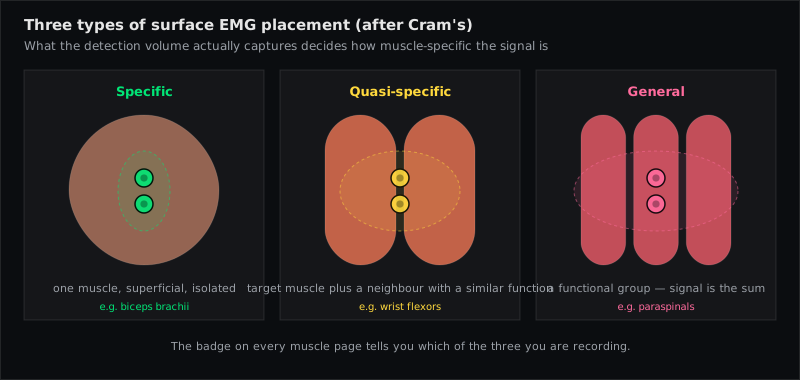
Surface EMG electrode placements come in three types, graded by how selectively they record: a specific placement records predominantly one muscle, a quasi-specific placement records a muscle together with neighbours it cannot be separated from, and a general (wide) placement deliberately records a whole region. The grading comes from Cram's electrode atlas (Criswell 2011) and is used on every EMG Guide muscle page. Which type you get is decided by anatomy and by inter-electrode distance — not by how carefully you palpate.
Why one muscle does not always equal one signal
Surface EMG can only "listen" so deep and so narrowly. Every bipolar pair records the summed activity of everything within its pickup volume — and that volume grows with the inter-electrode distance. Cram's rule of thumb: the depth and area of the recording are directly proportional to the spacing between the electrodes.
For a large, superficial, well-separated muscle such as the Biceps Brachii or the Gastrocnemius (Medial Head), a 20-mm pair over the belly records that muscle and little else. For the forearm flexors, the medial hamstrings or the hip adductors, the same pair inevitably records the neighbours too — the muscles are packed side by side under a few millimetres of skin, and their action potentials reach the electrodes by volume conduction regardless of how precisely the pair is centred.
Cram's therefore grades every atlas site rather than pretending each one is clean. That grading is the honest answer to a question practitioners ask constantly: "Am I really measuring this muscle?"
The three types
Specific placement
A closely spaced pair (SENIAM: 20 mm), parallel to the fibres, over or slightly lateral to the belly of a muscle that is superficial and has room around it. Cram's describes the Upper Trapezius site as the model case: on the ridge of the shoulder, slightly lateral to the centre of the belly, along the fibre direction.
What it records: predominantly the named muscle. Confidence comes from a functional test — the signal rises during the muscle's own action and stays quiet when neighbours contract.
Typical uses: recruitment patterns during movement, up-training and down-training of one muscle in biofeedback, side-to-side comparison of the same muscle.
Quasi-specific placement
The same electrode geometry — but over a muscle that is partly covered, very close to its neighbours, or thin enough that neighbouring activity is always part of the picture. Forearm flexors (Flexor Carpi Radialis, Flexor Digitorum Superficialis), the medial hamstrings (Semitendinosus + semimembranosus) and the hip adductors (Adductor Longus + magnus + gracilis) are Cram's standard examples.
What it records: the named muscle plus a share of the group. Cram's mentions one trick to sharpen it — for the finger flexors, place the pair on the distal part of the muscle where the tendon-side fibres are more exposed, so that finger movement (rather than wrist movement) dominates the trace.
What it means: interpret at group level. "The wrist flexors are active" is safe; "the flexor carpi radialis, but not the ulnaris" is not, unless a functional test proves it. Amplitude comparisons between two quasi-specific sites in the same group are unreliable.
General (wide) placement
Electrodes spaced far apart, often across several muscles or even limb-to-limb (Cram's mentions wrist-to-wrist and ankle-to-ankle leads in relaxation training). The wide spacing captures all muscle energy between the two active electrodes — and more.
What it records: a region. Basmajian's classic warning, quoted by Cram's: the wide frontal placement can pick up activity as far away as the first rib.
Typical uses: general relaxation training, screening for whole-region holding patterns. Not for muscle-specific statements of any kind.
Where SENIAM sits
SENIAM did not grade sites; it standardised them. Its 20-mm bipolar pair, fibre-parallel, between innervation zone and distal tendon at a defined landmark fraction is a specific-type geometry — applied to a catalogue of muscles that were selected in part because they can be recorded specifically. That is one reason the SENIAM set is as small as it is: 30-odd sites, 26 bilateral pairs on EMG Guide.
For muscles outside the catalogue EMG Guide uses published placements from Cram's and peer-reviewed studies. Many of those are quasi-specific by nature. The practice block on each muscle page states the type, so that a "quasi-specific" label is visible before you interpret the trace — not after.
What decides the type
Fridlund and Cacioppo's six elements for high-fidelity recordings (cited in Cram's Ch. 4) read like a checklist for reaching "specific":
- Proximity — the least tissue possible between electrodes and fibres.
- Orientation — parallel to the fibres; perpendicular pairs lose selectivity.
- Motor end-plate region avoided — straddling it lowers amplitude through differential cancellation; a little off-centre is better.
- Reproducible landmarks — sites you can find again next session.
- No obstruction — avoid skin folds, bony edges, positions that block movement.
- Electrode size and spacing chosen to minimise crosstalk from deep or neighbouring muscles.
You control 2–6. You do not control 1 — subcutaneous fat, muscle depth and how tightly the neighbours are packed are given. That is why a well-executed placement over the finger flexors is still quasi-specific, and why no amount of care turns a supraspinatus recording into a specific one (it lies under the trapezius; see which muscles cannot be recorded from the surface).
Practical consequences
- Verify, don't assume. Cram's: proof of specificity is that the signal responds to the movement ascribed to the muscle and not to contraction of the neighbours. Ask for both before you record.
- Match the claim to the type. Specific → statements about one muscle. Quasi-specific → statements about the group; use the trace for timing and pattern, be careful with amplitude ratios. General → statements about a region only.
- Compare like with like. Left–right comparison of the same quasi-specific site is fine (same neighbours on both sides). Comparing a quasi-specific site with a specific one, or two group members with each other, is not.
- Record several channels. Cram's recommends monitoring the synergist or antagonist alongside the target — the pattern across channels tells you more than one channel ever can, and it exposes crosstalk (a channel that mirrors its neighbour perfectly is suspicious).
- Note the type in your documentation together with position and spacing. It is part of the method.
Muscles referenced in this article
From placement to measurement
EMG Guide shows where the electrodes go. easyEMG with PicoBlue sensors visualises the signal live afterwards — SENIAM-conform, in real time, with a signal check on screen.
Frequently asked questions
Is a quasi-specific placement a bad placement?
No. It is the best available placement for a muscle that surface EMG cannot isolate. The mistake is not the placement but interpreting it as if it were specific.
Can a smaller inter-electrode distance make a quasi-specific site specific?
It increases selectivity and reduces crosstalk, so it helps — but it cannot overcome anatomy. Very small spacings also lower amplitude and make the recording more sensitive to exact position. SENIAM keeps 20 mm as the standard for comparability.
How do I know which type a muscle page describes?
Look at the "Recording notes" block on the muscle page: it states specific or quasi-specific per Cram's for that site, lists the crosstalk neighbours and the typical artifacts.
Does the type matter for biofeedback?
Yes. Up-training a specific site trains one muscle; up-training a quasi-specific site may train the group. For relaxation training a general placement is often exactly what you want.
Sources
- Criswell E. Cram's Introduction to Surface Electromyography. 2nd ed. Jones & Bartlett; 2011 — Ch. 1 (p. 5), Ch. 4 (pp. 70–72), Ch. 16 "Electrode Atlas Overview" (pp. 247–248).
- Fridlund AJ, Cacioppo JT. Guidelines for human electromyographic research. Psychophysiology. 1986;23:567–598.
- Hermens HJ et al. Development of recommendations for SEMG sensors and sensor placement procedures. J Electromyogr Kinesiol. 2000;10:361–374.
- Konrad P. The ABC of EMG. Noraxon; 2005 — p. 17.
Read next
EMG Electrode Placement — The Practical Guide
How to place surface EMG electrodes correctly — SENIAM rules, skin prep, 20 mm spacing, fibre direction, reference electrode, signal check and common mistakes.
Surface EMG Glossary
54 terms of surface electromyography explained in one sentence each — from amplitude cancellation to volume conduction. With sources (SENIAM, Cram's, Konrad).
Can I Measure This Muscle With Surface EMG?
Interactive check for 68 muscles: specific, quasi-specific or surface-recordable — or fine-wire only. Based on Cram's grades, SENIAM and the EMG Guide catalogue.
All articles →