Masseter (Masseter R)
The M. Masseter (Masseter) is a muscle of the head & neck region. It is responsible for jaw closure (elevation of the mandible), grinding during mastication. This muscle is not covered by the official SENIAM catalogue — placement is based on established clinical literature. This page documents the exact electrode position, anatomical attachments and a standardized test movement based on Hermens et al. (2000).
Jaw closure (elevation of the mandible), grinding during mastication
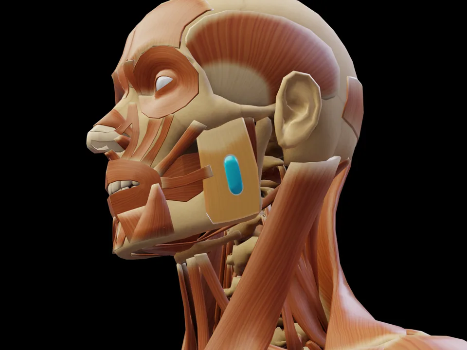
Preview — open the 3D viewer to rotate the model and check neighbouring muscles.
View in 3D Viewer
Over the muscle belly at the angle of the jaw — palpate while the person clenches the teeth and place the electrodes over the clearly protruding belly
Orientation: Along the muscle fibres, 2 cm inter-electrode distance; place the pairs symmetrically when comparing sides
Criswell E. Cram's Introduction to Surface Electromyography. 2nd ed. Jones & Bartlett Learning; 2011. ISBN 978-0-7637-3274-5.
Criswell (2011) — Cram's Introduction to Surface Electromyography, 2nd edition — is the standard textbook for surface EMG electrode placement beyond the SENIAM catalogue. Its atlas section (Part III) describes, for each site, palpation, electrode location with 2 cm spacing parallel to the fibres, a behavioural test and the neighbouring muscles from which crosstalk is to be expected. Cram's classifies the position used here as "quasi-specific" — the muscle cannot be fully separated from its neighbours at the surface. The position is established in practice but less validated than a SENIAM recommendation.
Based on SENIAM recommendations (Hermens et al. 2000). Not an official SENIAM product. See all references → · How electrodes are placed (guide) · Glossary
Innervation after Perotto AO, Anatomical Guide for the Electromyographer, 5th ed. 2011 — an anatomical reference, not an electrode placement recommendation.
Clench the teeth firmly
This muscle has no SENIAM-standardized electrode position. Placement should be based on anatomical knowledge.
The electrode is placed — what next? easyEMG visualises muscle activity in real time, with SENIAM-compliant placement guidance, up to 4 channels at once and biofeedback training right afterwards.
In therapy, this muscle is usually about tone regulation and consciously relaxing below a threshold.
Over the muscle belly at the angle of the jaw — palpate while the person clenches the teeth and place the electrodes over the clearly protruding belly. Orientation: Along the muscle fibres, 2 cm inter-electrode distance; place the pairs symmetrically when comparing sides. Not covered by SENIAM — placement per Criswell 2011.
Clench the teeth firmly. It serves as a signal check before recording: with correct electrode placement the EMG amplitude rises clearly during this movement.
No. The M. Masseter is not included in the official SENIAM catalogue. The electrode position documented here follows the established protocol per Criswell 2011.
Interactive SENIAM electrode placement — free, instant access, no sign-up required.
Open 3D Viewer →49 muscle protocols (SENIAM + peer-reviewed) on 85 pages, print-ready. Free via email.
Professional EMG biofeedback software for PicoBlue sensors. Real-time visualization, 5 training modes, PDF reports.