Recording notes & source
per Criswell 2011, site „Hip Adductor (Adductor Longus/Gracilis)“, p. 367
- Placement type
- Quasi-specific site Neighbouring muscles contribute noticeably to the signal — allow for crosstalk.
- Signal check
- Press the knees together (adduction); walking.
- Crosstalk from
- adductor longus (inseparable at the surface) · rectus femoris · vastus medialis · adductor magnus
Source:
Criswell 2011
Criswell E. Cram's Introduction to Surface Electromyography. 2nd ed. Jones & Bartlett Learning; 2011. ISBN 978-0-7637-3274-5.
Methodological background of the source
Criswell (2011) — Cram's Introduction to Surface Electromyography, 2nd edition — is the standard textbook for surface EMG electrode placement beyond the SENIAM catalogue. Its atlas section (Part III) describes, for each site, palpation, electrode location with 2 cm spacing parallel to the fibres, a behavioural test and the neighbouring muscles from which crosstalk is to be expected. Cram's classifies the position used here as "quasi-specific" — the muscle cannot be fully separated from its neighbours at the surface. The position is established in practice but less validated than a SENIAM recommendation.
Based on SENIAM recommendations (Hermens et al. 2000). Not an official SENIAM product. See all references →
· How electrodes are placed (guide)
· Glossary
All 49 muscle protocols as a PDF — free
Print-ready for the lab or clinic: 79 pages of protocols (SENIAM + peer-reviewed), plus the SENIAM table, a placement checklist and a sensor comparison sheet as A4 sheets.
Anatomy & Function
The M. Gracilis (Gracilis) is a muscle of the hip region. It is responsible for hip adduction, knee flexion and internal rotation. This muscle is not covered by the official SENIAM catalogue — placement is based on established clinical literature. This page documents the exact electrode position, anatomical attachments and a standardized test movement based on Hermens et al. (2000).
Function
Hip adduction, knee flexion and internal rotation
Origin
Inferior ramus of the pubis, near the symphysis
Insertion
Pes anserinus (proximal medial tibial surface)
Innervation
Obturator nerve, L2–L4
Test Movement
Thigh adduction (pressing the knees together) against resistance
This muscle has no SENIAM-standardized electrode position. Placement should be based on anatomical knowledge.
MVC reference test (Konrad 2005)
- Position
- Supine or seated with flexed legs; a big, stiff roll cushion is pressed between the knees.
- Fixation
- The cushion itself provides the rigid resistance; pelvis stable.
- Resistance
- Isometric adduction against the cushion.
Konrad tests the adductors as one group ("Mm. adductores"); surface EMG does not separate longus, magnus and gracilis.
Konrad P. The ABC of EMG. Noraxon; 2005, p. 32. Konrad describes this test at group level; it applies to all muscles of the group. · Why and how to normalise to MVC
Measure M. Gracilis activity live
The electrode is placed — what next? easyEMG visualises muscle activity in real time, with SENIAM-compliant placement guidance, up to 4 channels at once and biofeedback training right afterwards.
In rehabilitation, the Gracilis is typically recorded to track progress and retrain activation.
Frequently Asked Questions
Where do I place the EMG electrode on the M. Gracilis?
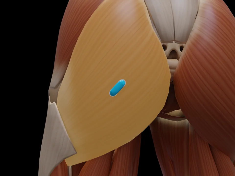
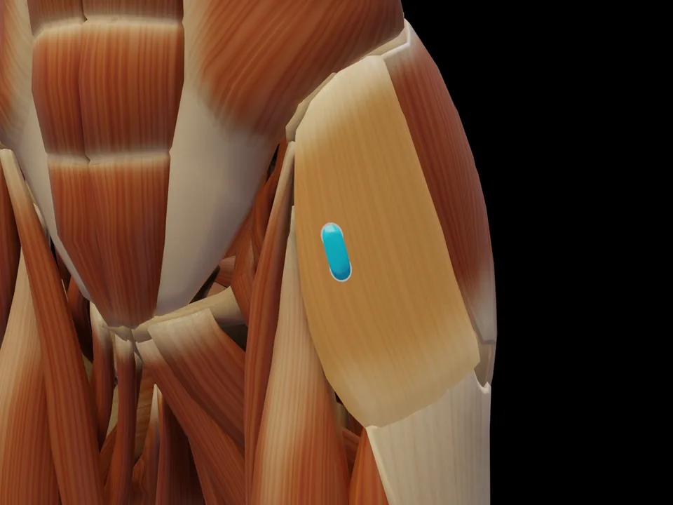
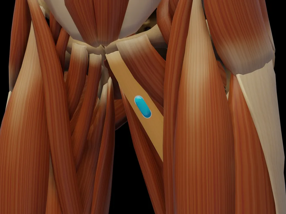
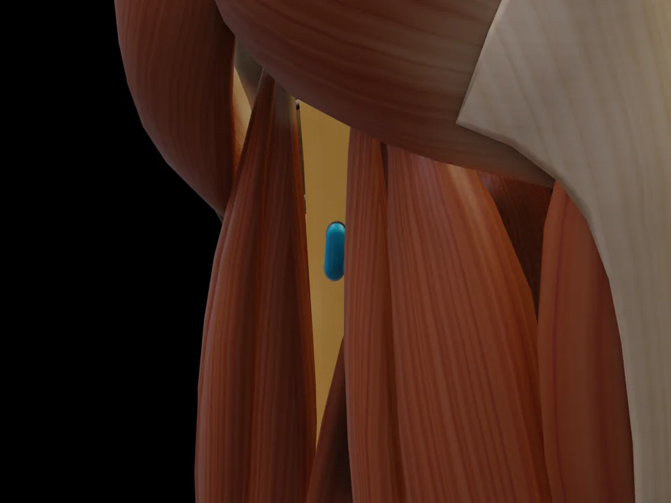
Medial thigh, approx. 4 cm distal to the pubis; palpate the area during isometric adduction. Note: hip-adductor site per Criswell — cannot be separated from the adductor longus at the surface. Orientation: Oblique along the muscle course, 2 cm inter-electrode distance; avoid the anterior midline of the thigh (crosstalk from rectus femoris). Not covered by SENIAM — placement per Criswell 2011.
Which test movement activates the M. Gracilis?
Thigh adduction (pressing the knees together) against resistance. It serves as a signal check before recording: with correct electrode placement the EMG amplitude rises clearly during this movement.
Is the M. Gracilis a SENIAM-recommended muscle?
No. The M. Gracilis is not included in the official SENIAM catalogue. The electrode position documented here follows the established protocol per Criswell 2011.
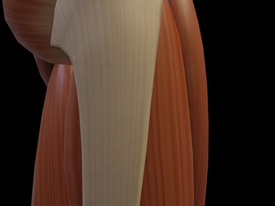 Preview — open the 3D viewer to rotate the model and check neighbouring muscles.
View in 3D Viewer
Preview — open the 3D viewer to rotate the model and check neighbouring muscles.
View in 3D Viewer