Which Muscles Cannot Be Measured With Surface EMG — and Why
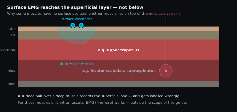
Surface EMG can only record muscles that lie directly under the skin. A muscle that is covered by another muscle — the supraspinatus under the trapezius, the iliopsoas behind the abdominal wall, the tibialis posterior under the soleus — cannot be isolated with surface electrodes: whatever the pair picks up is dominated by the muscle on top. Such muscles are recorded with intramuscular fine-wire or needle electrodes, and that is why EMG Guide deliberately does not list them, even though they are clinically important.
Quick answer for a specific muscle: use the interactive check Can I measure this muscle with surface EMG?
The physics in one paragraph
Action potentials travel through tissue by volume conduction, and tissue attenuates them with distance — muscle, fat and skin act as a low-pass filter. A bipolar surface pair with 20 mm spacing "sees" roughly as deep as it is wide (Cram's: depth and area of the recording are proportional to the inter-electrode distance). If a second, active muscle lies between the target and the skin, its potentials arrive larger, sharper and earlier than anything from below. Cram's is explicit: it is impossible to state that such a recording is primarily from the deeper muscle, because it can easily be contaminated by the superficial layer. Increasing the spacing to "reach deeper" only widens the pickup and adds more of the surface muscle.
Muscles that surface EMG cannot isolate
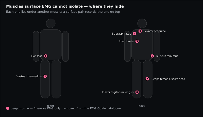
The muscles below were part of earlier EMG Guide versions and were removed after checking the sources behind their supposed surface positions. In every case the primary literature uses intramuscular electrodes, or the position turned out to be a placement over the covering muscle.
| Muscle | Lies under | What a surface pair actually records | How the literature records it |
|---|---|---|---|
| Supraspinatus | Upper trapezius | Trapezius (upper part) | Intramuscular (e.g. Boettcher et al. 2008); Perotto: needle through the trapezius |
| Levator scapulae | Upper trapezius, sternocleidomastoid | Trapezius | Fine-wire (Castelein et al. 2016); Perotto |
| Rhomboid major/minor | Middle trapezius | Trapezius (middle part) | Fine-wire (Castelein et al. 2016); Perotto |
| Iliopsoas | Abdominal wall, inguinal region | Abdominal muscles, sartorius, adductors | Fine-wire; surface attempts in the femoral triangle are unreliable and unsafe to interpret |
| Gluteus minimus | Gluteus medius | Gluteus medius | Fine-wire |
| Vastus intermedius | Rectus femoris | Rectus femoris | Fine-wire |
| Biceps femoris short head | Long head | Long head | Fine-wire |
| Flexor digitorum longus | Soleus / gastrocnemius | Soleus | Fine-wire |
Never part of the catalogue, for the same reason: tibialis posterior (under the soleus — Konrad shows a fine-wire tibialis posterior trace as his standard example), pectoralis minor (under the pectoralis major; fine-wire in Castelein et al. 2016), the deep neck flexors (longus colli/capitis, behind the sternocleidomastoid and pharynx), subscapularis, popliteus, the diaphragm, the intrinsic hand muscles under the palmar aponeurosis, and most pelvic-floor muscles (which need probes rather than skin electrodes). The multifidus is listed — but its L5 surface site is quasi-specific: a mix with the longissimus, as the muscle page says.
What you can do instead
Record the covering muscle and say so. A pair over the upper trapezius records the upper trapezius. That is a valid, specific measurement — as long as it is labelled as such. Cram's atlas grades every site by specificity (see specific, quasi-specific, general); the honest grade for a "supraspinatus" surface site is trapezius.
Use a functional test to see what you have. Cram's rule for specificity: the trace must respond to the movement ascribed to the target and stay quiet during contraction of the neighbours. Ask for shoulder abduction with the trapezius relaxed and watch whether the "supraspinatus" channel is really independent of shrugging. It will not be.
Study the accessible synergists. For the rotator cuff, Infraspinatus is superficial below the scapular spine and records well; for scapular control, Middle Trapezius, the lower trapezius and Serratus Anterior are accessible; for the hip, Gluteus Medius is; for the knee extensors, Rectus Femoris and the vasti; for the hamstrings, the long head of the Biceps Femoris (Long Head); for the plantar flexors, Soleus and the gastrocnemii. Their patterns tell you a great deal about the deep muscle's job even without recording it.
Refer to fine-wire EMG when the deep muscle itself is the question. Konrad: for deeper muscles covered by surface muscles or bone, fine-wire or needle electrodes are inevitable. It is a research and clinical-neurophysiology tool, not a biofeedback tool — but for a research question about the iliopsoas or the tibialis posterior there is no surface shortcut.
Why EMG Guide removed these muscles
In 2026 the catalogue was reduced to 106 muscles (the masseter pair has been added since — 108 today). Each removal followed the same check: does the cited source actually record this muscle with surface electrodes at the stated position? For the muscles above the answer was no — the source used intramuscular electrodes, described a different muscle, or gave no position at all. Keeping a "position" that in reality records the trapezius would mislabel every measurement made with it. The old URLs redirect to the respective body-region overview; the sources behind every remaining position are listed on the references page.
Muscles referenced in this article
From placement to measurement
EMG Guide shows where the electrodes go. easyEMG with PicoBlue sensors visualises the signal live afterwards — SENIAM-conform, in real time, with a signal check on screen.
Frequently asked questions
Can I measure the iliopsoas with surface EMG?
Not in a way that can be attributed to the iliopsoas. Surface pairs in the femoral triangle sit over the sartorius, adductors and abdominal wall and record those. Studies of the iliopsoas use fine-wire electrodes.
Why is the supraspinatus not on EMG Guide even though studies "measure" it?
Because the studies that measure it use intramuscular electrodes (e.g. Boettcher et al. 2008); the surface electrodes in those same studies sit on the trapezius and deltoid. A surface pair over the supraspinous fossa records the upper trapezius.
Is there any deep muscle that surface EMG can reach?
Only where the covering layer is thin, inactive or absent for the task — the multifidus at L5 is the usual example, and even there the recording is quasi-specific (a mix with the longissimus). If the covering muscle is active during the movement you study, the deep muscle cannot be separated.
Does a smaller or larger electrode spacing help?
Smaller spacing makes the recording more selective for the surface layer; larger spacing widens the pickup and adds more of everything. Neither lets you skip a muscle that lies on top.
What should I write in a report when I recorded over a covered muscle?
Name the muscle that is actually under the electrodes (e.g. "upper trapezius, position over the supraspinous fossa"), state that the deep target cannot be isolated with surface electrodes, and interpret at group level.
Sources
- Perotto AO. Anatomical Guide for the Electromyographer. 5th ed. Charles C Thomas; 2011 — levator scapulae, rhomboids, supraspinatus (needle through the trapezius), iliopsoas, tibialis posterior.
- Criswell E. Cram's Introduction to Surface Electromyography. 2nd ed. 2011 — Ch. 1 (advantages and limitations of SEMG, p. 5), Ch. 16 (pp. 247–248).
- Boettcher CE, Ginn KA, Cathers I. Standard maximum isometric voluntary contraction tests for normalizing shoulder muscle EMG. J Orthop Res. 2008;26:1591–1597 (intramuscular electrodes for supraspinatus).
- Castelein B et al. Serratus anterior or pectoralis minor: which muscle has the upper hand during protraction exercises? Man Ther. 2016;22:158–164 (fine-wire for levator scapulae, rhomboid major, pectoralis minor).
- Konrad P. The ABC of EMG. Noraxon; 2005 — p. 15–16 (surface vs. fine-wire electrodes).
Read next
EMG Electrode Placement — The Practical Guide
How to place surface EMG electrodes correctly — SENIAM rules, skin prep, 20 mm spacing, fibre direction, reference electrode, signal check and common mistakes.
Specific, Quasi-Specific, General — The Three Types of Surface EMG Placement
Why there is no single correct electrode position — Cram's three placement types, what each records, where SENIAM fits, and what it means for interpretation.
Surface EMG Glossary
54 terms of surface electromyography explained in one sentence each — from amplitude cancellation to volume conduction. With sources (SENIAM, Cram's, Konrad).
All articles →