Recording notes & source
per Criswell 2011, site „Hip Flexor (Sartorius)“, p. 361
- Placement type
- Quasi-specific site Neighbouring muscles contribute noticeably to the signal — allow for crosstalk.
- Signal check
- Supine: flex and externally rotate the thigh while flexing the knee.
- Crosstalk from
- rectus femoris · iliopsoas · adductor magnus
Source:
Criswell 2011
Criswell E. Cram's Introduction to Surface Electromyography. 2nd ed. Jones & Bartlett Learning; 2011. ISBN 978-0-7637-3274-5.
Methodological background of the source
Criswell (2011) — Cram's Introduction to Surface Electromyography, 2nd edition — is the standard textbook for surface EMG electrode placement beyond the SENIAM catalogue. Its atlas section (Part III) describes, for each site, palpation, electrode location with 2 cm spacing parallel to the fibres, a behavioural test and the neighbouring muscles from which crosstalk is to be expected. Cram's classifies the position used here as "quasi-specific" — the muscle cannot be fully separated from its neighbours at the surface. The position is established in practice but less validated than a SENIAM recommendation.
Based on SENIAM recommendations (Hermens et al. 2000). Not an official SENIAM product. See all references →
· How electrodes are placed (guide)
· Glossary
All 49 muscle protocols as a PDF — free
Print-ready for the lab or clinic: 79 pages of protocols (SENIAM + peer-reviewed), plus the SENIAM table, a placement checklist and a sensor comparison sheet as A4 sheets.
Anatomy & Function
The M. Sartorius (Sartorius) is a muscle of the thigh anterior region. It is responsible for hip flexion, abduction and external rotation; knee flexion and internal rotation (longest muscle of the body). This muscle is not covered by the official SENIAM catalogue — placement is based on established clinical literature. This page documents the exact electrode position, anatomical attachments and a standardized test movement based on Hermens et al. (2000).
Function
Hip flexion, abduction and external rotation; knee flexion and internal rotation (longest muscle of the body)
Origin
Anterior superior iliac spine (ASIS)
Insertion
Pes anserinus (proximal medial tibial surface)
Innervation
Femoral nerve, L2–L4
Test Movement
Supine: flex and externally rotate the thigh while flexing the knee, against resistance
This muscle has no SENIAM-standardized electrode position. Placement should be based on anatomical knowledge.
Measure M. Sartorius activity live
The electrode is placed — what next? easyEMG visualises muscle activity in real time, with SENIAM-compliant placement guidance, up to 4 channels at once and biofeedback training right afterwards.
In rehabilitation, the Sartorius is typically recorded to track progress and retrain activation.
Frequently Asked Questions
Where do I place the EMG electrode on the M. Sartorius?
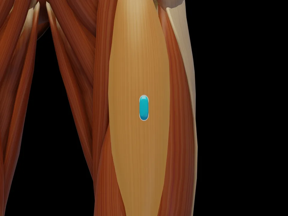
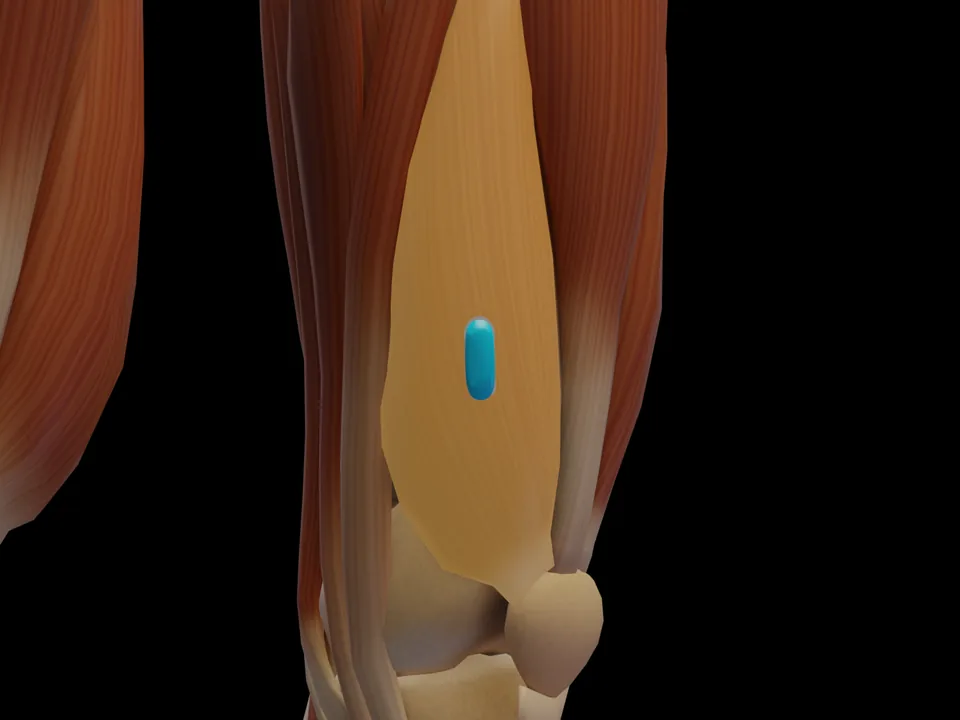
Proximal anterior thigh, approx. 4 cm distal to the anterior superior iliac spine; stay proximal to minimise crosstalk from the rectus femoris. Orientation: Oblique along the muscle course (from the ASIS medially and distally), 2 cm inter-electrode distance. Not covered by SENIAM — placement per Criswell 2011.
Which test movement activates the M. Sartorius?
Supine: flex and externally rotate the thigh while flexing the knee, against resistance. It serves as a signal check before recording: with correct electrode placement the EMG amplitude rises clearly during this movement.
Is the M. Sartorius a SENIAM-recommended muscle?
No. The M. Sartorius is not included in the official SENIAM catalogue. The electrode position documented here follows the established protocol per Criswell 2011.
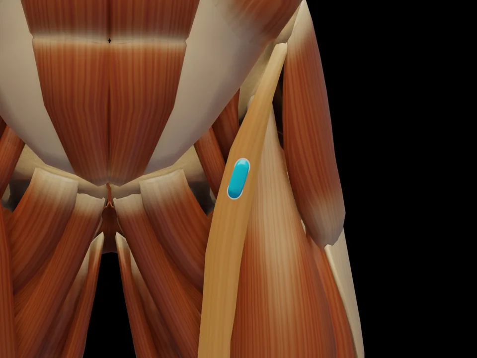 Preview — open the 3D viewer to rotate the model and check neighbouring muscles.
View in 3D Viewer
Preview — open the 3D viewer to rotate the model and check neighbouring muscles.
View in 3D Viewer