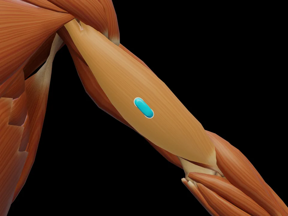
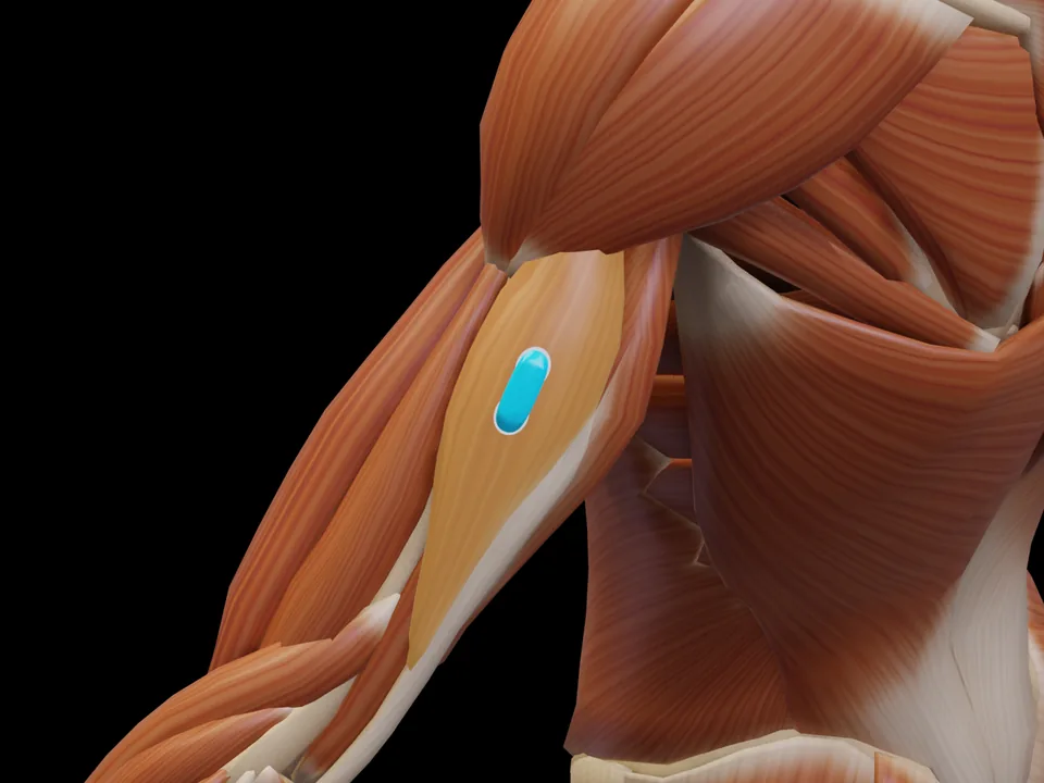
In the distal third of the upper arm where the muscle becomes superficial - two to three fingerbreadths lateral to the line from acromion to cubital fossa, clear of biceps and triceps
OrientationAlong the muscle fibres, 2 cm inter-electrode distance
Date S, Kurumadani H, Nakashima Y, Ishii Y, Ueda A, Kurauchi K, Sunagawa T. Brachialis muscle activity can be measured with surface electromyography: a comparative study using surface and fine-wire electrodes. Front Physiol. 2021;12:809422.
Methodological background of the source
Date et al. (2021) used ultrasound to verify whether the brachialis can be recorded with surface electrodes at all, comparing the signal directly against a fine-wire recording in the same muscle. The position shown here marks the distal region where the muscle emerges beside the biceps — only there can it be recorded without crosstalk from biceps and triceps.
Print-ready for the lab or clinic: 79 pages of protocols (SENIAM + peer-reviewed), plus the SENIAM table, a placement checklist and a sensor comparison sheet as A4 sheets.
Anatomy & Function
The M. Brachialis (Brachialis) is a muscle of the upper arm region. It is responsible for elbow flexion (strongest elbow flexor, independent of pronation/supination). This muscle is not covered by the official SENIAM catalogue — placement is based on established clinical literature. This page documents the exact electrode position, anatomical attachments and a standardized test movement based on Hermens et al. (2000).
FunctionElbow flexion (strongest elbow flexor, independent of pronation/supination)OriginDistal half of the anterior humerusInsertionUlnar tuberosity, coronoid processInnervationMusculocutaneous nerve (lateral cord), C5–C6; plus a small branch of the radial nerve
Innervation after Perotto AO, Anatomical Guide for the Electromyographer, 5th ed. 2011 — an anatomical reference, not an electrode placement recommendation.
Test Movement
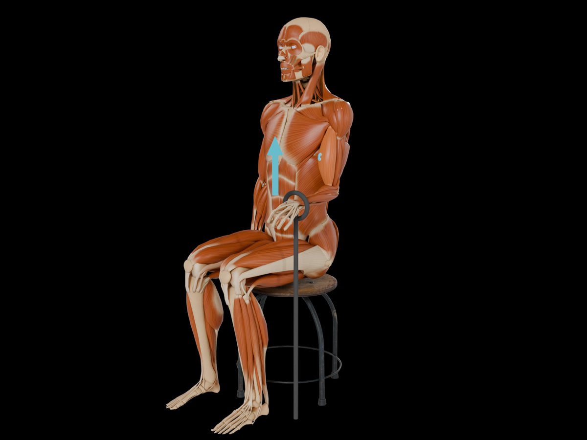
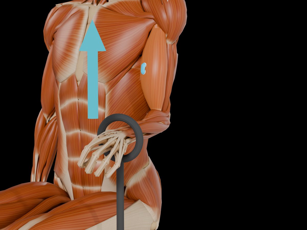
Elbow flexion against resistance with pronated forearm
This muscle has no SENIAM-standardized electrode position. Placement should be based on anatomical knowledge.
Illustration shows the left side; the set-up is mirrored for the right side.
MVC reference test (Konrad 2005)
Position
Seated or kneeling in front of a bench, elbow at about 90°, forearm supinated.
Fixation
Very stable fixation of elbow and trunk; the upper arm stays vertical at the side.
Resistance
Rigid resistance under the wrist (strap to the floor, bar or manual), elbow flexion against it.
Control exercise
Use the latissimus dorsi test as a control exercise.
Without elbow and trunk fixation the effort leaks into the shoulder and the biceps never reaches its maximum.
Konrad P. The ABC of EMG. Noraxon; 2005, p. 31. Konrad describes this test at group level; it applies to all muscles of the group. · Why and how to normalise to MVC
Measure M. Brachialis activity live
The electrode is placed — what next? easyEMG visualises muscle activity in real time, with SENIAM-compliant placement guidance, up to 4 channels at once and biofeedback training right afterwards.
In rehabilitation, the Brachialis is typically recorded to track progress and retrain activation.
Where do I place the EMG electrode on the M. Brachialis?
In the distal third of the upper arm where the muscle becomes superficial - two to three fingerbreadths lateral to the line from acromion to cubital fossa, clear of biceps and triceps. Orientation: Along the muscle fibres, 2 cm inter-electrode distance. Not covered by SENIAM — placement per Date et al. 2021.
Which test movement activates the M. Brachialis?
Elbow flexion against resistance with pronated forearm. It serves as a signal check before recording: with correct electrode placement the EMG amplitude rises clearly during this movement.
Is the M. Brachialis a SENIAM-recommended muscle?
No. The M. Brachialis is not included in the official SENIAM catalogue. The electrode position documented here follows the established protocol per Date et al. 2021.
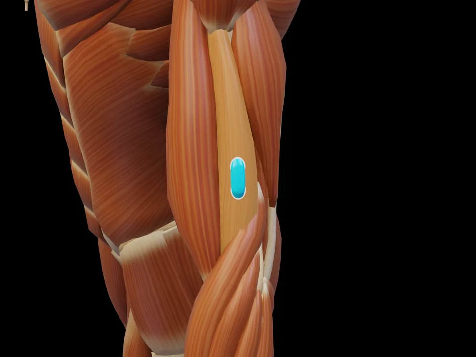 Preview — open the 3D viewer to rotate the model and check neighbouring muscles.
View in 3D Viewer
Preview — open the 3D viewer to rotate the model and check neighbouring muscles.
View in 3D Viewer