Hermens HJ, Freriks B, Disselhorst-Klug C, Rau G. Development of recommendations for SEMG sensors and sensor placement procedures. J Electromyogr Kinesiol. 2000;10(5):361-74.
Print-ready for the lab or clinic: 79 pages of protocols (SENIAM + peer-reviewed), plus the SENIAM table, a placement checklist and a sensor comparison sheet as A4 sheets.
Anatomy & Function
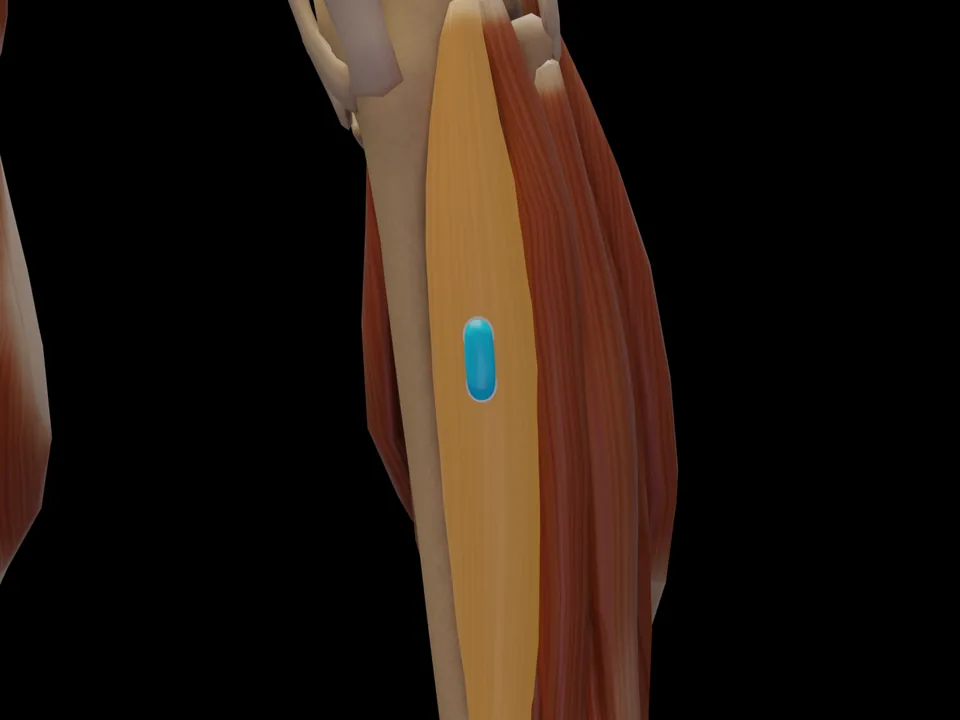
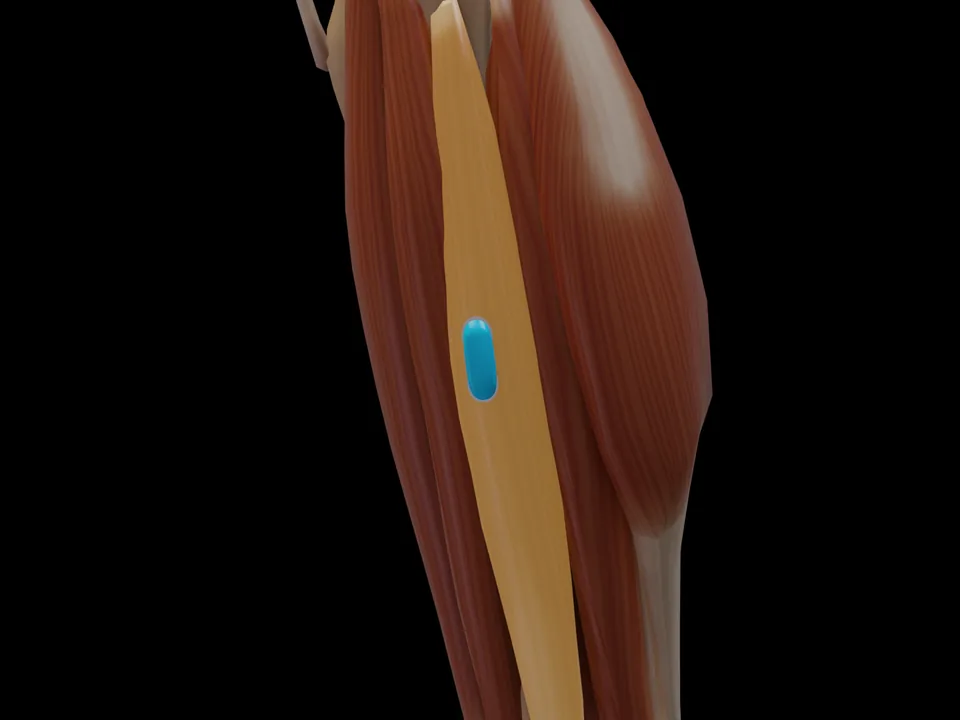
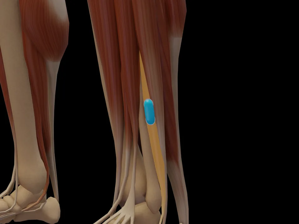
The M. Gastrocnemius Caput Mediale (Gastrocnemius (Medial Head)) is a muscle of the lower leg region. It is responsible for ankle plantar flexion, knee flexion (biarticular), push-off during walking/running. This muscle is one of 26 with official SENIAM-recommended surface EMG electrode placement. This page documents the exact electrode position, anatomical attachments and a standardized test movement based on Hermens et al. (2000).
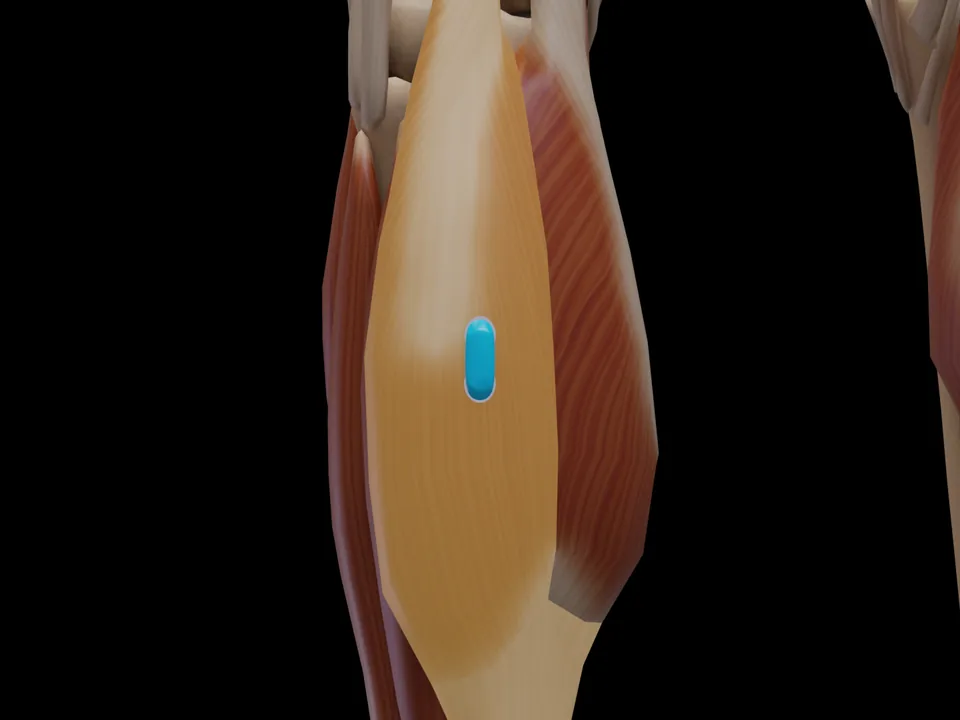
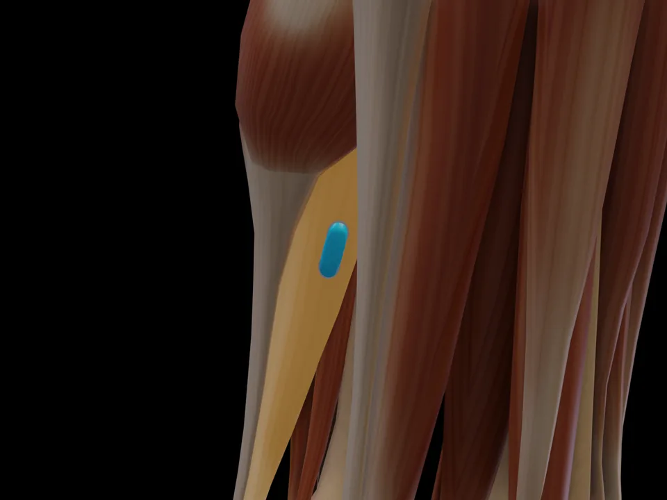
FunctionAnkle plantar flexion, knee flexion (biarticular), push-off during walking/runningOriginMedial epicondyle of the femur (posterior)InsertionCalcaneus (via Achilles tendon / tendo calcaneus)InnervationTibial nerve, S1–S2
Innervation after Perotto AO, Anatomical Guide for the Electromyographer, 5th ed. 2011 — an anatomical reference, not an electrode placement recommendation.
Clinical applications (SENIAM)
Gait analysis and foot drop assessment
Achilles tendinopathy monitoring
Proprioceptive training after ankle injury
Test Movement
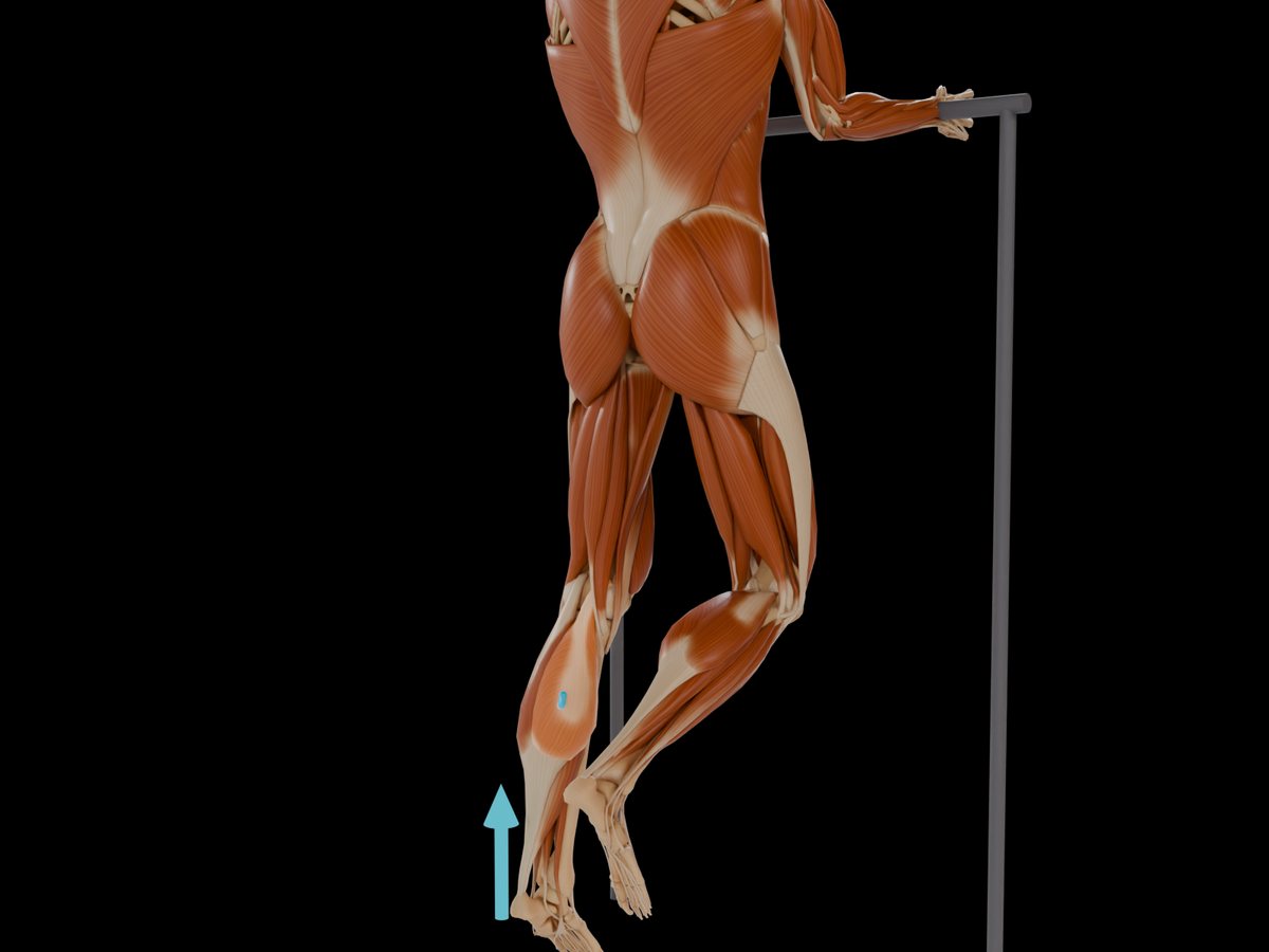
Heel raise (single leg) or plantar flexion against resistance with extended knee
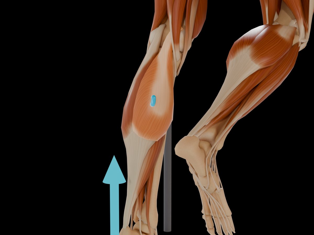
Illustration shows the left side; the set-up is mirrored for the right side.
MVC reference test (Konrad 2005)
Position
Unilateral plantar flexion at a 90° ankle position with the knee extended; standing single-leg heel raise as a field alternative.
Fixation
Very rigid (machine) resistance against the fixed hip; standing: hands on a rail for balance only.
Resistance
Fixed footplate or body weight plus load, plantar flexion against it.
Control exercise
The soleus test (knee flexed) is the check exercise.
Konrad: one of the strongest human muscle groups — the triceps surae needs very rigid resistance, otherwise the maximum is not reached.
Konrad P. The ABC of EMG. Noraxon; 2005, p. 32. Konrad describes this test at group level; it applies to all muscles of the group. · Why and how to normalise to MVC
Measure M. Gastrocnemius Caput Mediale activity live
The electrode is placed — what next? easyEMG visualises muscle activity in real time, with SENIAM-compliant placement guidance, up to 4 channels at once and biofeedback training right afterwards.
In screening, this muscle is usually recorded together with its synergists — running several channels in parallel saves the most time.
Where do I place the EMG electrode on the M. Gastrocnemius Caput Mediale?
Most proximal part of the medial muscle belly. Orientation: Parallel to the longitudinal axis of the lower leg. Based on the official SENIAM recommendations (Hermens et al. 2000).
Which test movement activates the M. Gastrocnemius Caput Mediale?
Heel raise (single leg) or plantar flexion against resistance with extended knee. This movement is part of the SENIAM protocol for standardized contraction verification.
Is the M. Gastrocnemius Caput Mediale a SENIAM-recommended muscle?
Yes. The M. Gastrocnemius Caput Mediale is one of 26 muscles for which the SENIAM project has published official surface EMG electrode placement recommendations.
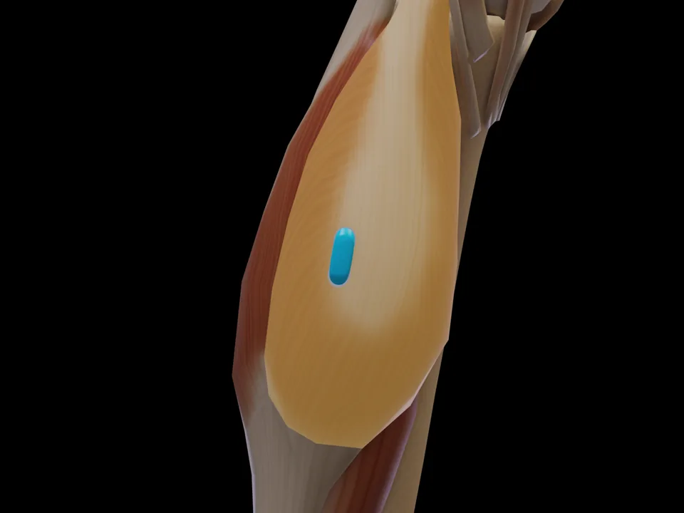 Preview — open the 3D viewer to rotate the model and check neighbouring muscles.
View in 3D Viewer
Preview — open the 3D viewer to rotate the model and check neighbouring muscles.
View in 3D Viewer