EMG Parameters — Which Number Answers Which Question
A surface EMG recording can answer six kinds of question — is the muscle on or off, is it more or less active than in another condition, when does it fire relative to others, how much of its capacity does it use, does it fatigue, and how does it coordinate with its neighbours (Konrad's six analysis levels). Each question has its own parameter: onset/offset thresholds, mean amplitude, time-to-peak and firing order, %MVC, median-frequency slope, co-activation indices. Choosing the parameter before the recording — not after — is what makes an EMG measurement interpretable.
Why start with the question
Konrad's analysis chapter is built around a simple observation: the parameters are cheap, the question is expensive. Software will compute mean, peak, area, median frequency and onsets for every channel in a second — but a mean amplitude cannot answer a timing question, a peak cannot answer a fatigue question, and a single-channel value cannot answer a coordination question. Cram's makes the same point from the clinical side (Ch. 8): decide what you want to know, then choose the placement, the task and the number.
Every parameter below assumes the processing chain from raw EMG to RMS and, where amplitudes are compared across sessions or people, normalisation.
Level 1 — On or off?
Question: Is the muscle active at all during this task, and when?
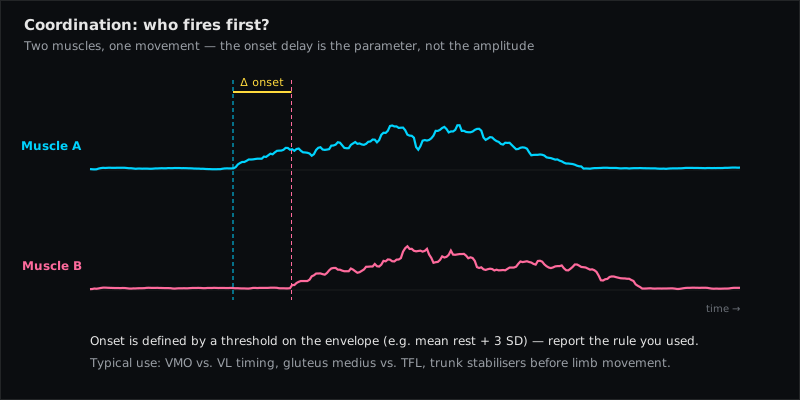
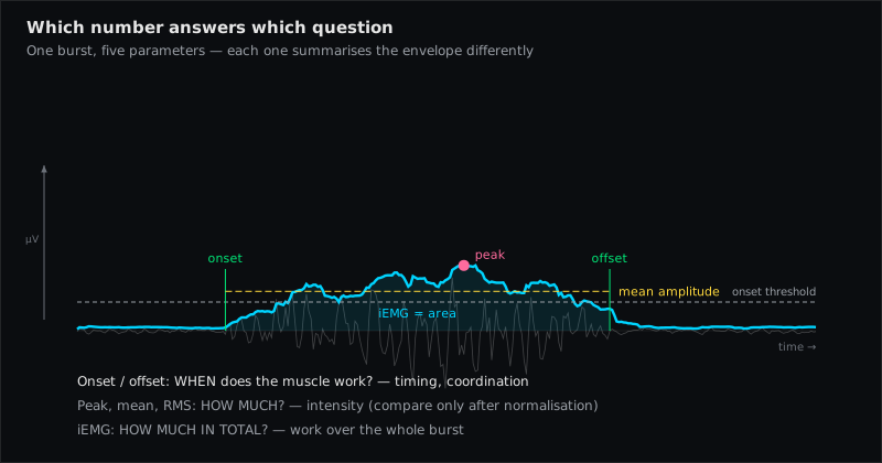
Parameter: onset and offset, defined by a threshold on the envelope. Konrad lists three ways to set it:
- Multiple of baseline SD — typically 2–3 standard deviations of the resting baseline, plus a minimum duration (e.g. 50 ms) above threshold so single spikes do not trigger. Popular, but the baseline SD varies between trials and people; with very quiet amplifiers the factor may need to rise to 8 or more.
- Percentage of local peak — e.g. 5 % of the peak within the analysis window; more stable, independent of baseline noise.
- Fixed value — a microvolt level, or better a % MVC level in normalised data.
Whatever the rule: check every onset graphically. Hodges & Bui showed how much the detected onset moves with method and window — a timing conclusion is only as good as its threshold.
Level 2 — More or less?
Question: Is the muscle more active in condition A than in B — left vs. right, before vs. after, exercise 1 vs. exercise 2?
Parameter: the mean amplitude of the envelope over a defined interval — Konrad's "most important EMG calculation": robust, least sensitive to interval length, best for comparisons. Peak is too variable on single trials; use the average peak (mean of the ten highest values) or peaks of averaged curves. Area (iEMG) only at equal interval lengths.
Rules: same electrodes, same task, same window; within one session raw µV are acceptable for left–right; across sessions or people normalise. And remember Cram's warning: more microvolts is not more force.
Level 3 — When? (timing)
Question: In which order do the muscles fire, how long after a stimulus, when is the peak?
Parameters: time to peak (from contraction start or event to peak of the envelope), firing order across channels (onset ranking), onset pattern diagrams (on/off bars per muscle across a movement cycle — the classic gait representation), and reaction time after an external trigger.
Rules: short smoothing windows or zero-phase filters (every moving window delays onsets), a synchronised event marker (foot switch, goniometer, video), and identical threshold rules for all channels. Typical clinical questions: does the Vastus Medialis start before the Vastus Lateralis? Does the Gluteus Maximus fire before the Biceps Femoris (Long Head) in hip extension? Does the Multifidus switch on in a feed-forward manner before the arm moves?
Level 4 — How much?
Question: How much of its capacity is the muscle using?
Parameter: mean amplitude expressed as % MVC (or another reference — see the normalisation article). Only normalised data can say "the Upper Trapezius worked at 15 % of maximum during this typing task". Konrad's Input % — each channel's mean as a share of the summed means — answers the related question "how is the effort distributed across these muscles?"
Rules: the reference contraction must be valid (see MVC test rules); values above 100 % occur in dynamic tasks and are a reason to check the reference, not to clip.
Level 5 — Does it fatigue?
Question: Does the muscle show signs of local fatigue during a sustained effort?
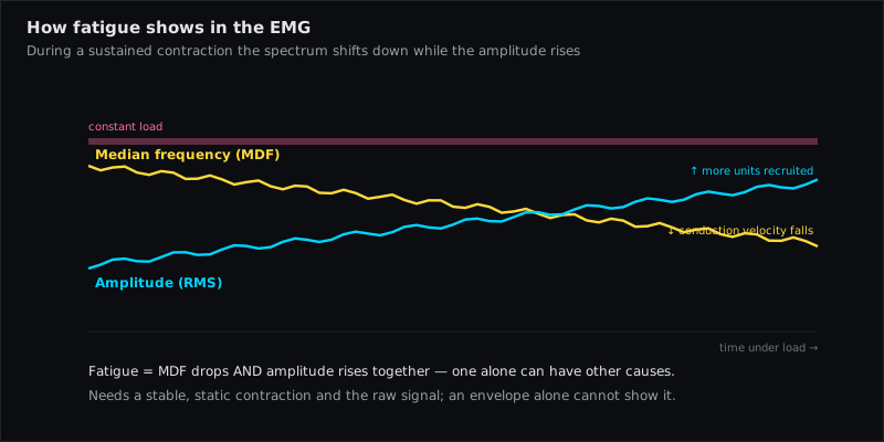
Parameters: in a static, submaximal contraction at constant load and fixed joint angle, two things happen over time (Konrad, p. 50): the amplitude rises (additional motor units are recruited) and the mean and median frequency of the power spectrum fall (conduction velocity slows; the spectrum shifts left). The slope of median frequency over time — with its intercept — is the classic muscle fatigue index (De Luca). It appears before force actually declines.
Rules: the spectral method needs stationarity — a constant, static contraction. In dynamic tasks (strength training) use the amplitude rise instead. Watch for the opposite pattern (frequency up, amplitude down), which Konrad attributes to load shifting to synergists or reduced antagonist co-activation. Applications: back-endurance tests (multifidus and erector spinae in low-back pain), evaluating training effects.
Level 6 — Coordination
Question: Do the muscles around a joint or along a chain work together appropriately?
Parameters: all of the above, but on at least two channels and with explicit criteria. Konrad's examples of "good" coordination: symmetrical innervation of synergists (the vasti), synchronised firing order within a chain, feed-forward activation of stabilisers, and antagonist co-innervation that is low and late. Co-activation is quantified from the overlap of agonist and antagonist envelopes; ratios such as VMO/VL or upper/lower trapezius are the clinical shorthand.
Rules: define the criterion before recording; record synergists and antagonists together; watch for crosstalk masquerading as co-activation (a channel that mirrors its neighbour perfectly is suspicious).
The map
| Question | Parameter | Needs | Typical trap |
|---|---|---|---|
| On/off? | onset/offset by threshold | short window, defined threshold, visual check | threshold rule changes the answer |
| More/less? | mean amplitude of envelope | same setup; normalise across sessions/people | reading µV as force |
| When? | time to peak, firing order, onset pattern | event marker, zero-phase smoothing | window lag shifts onsets |
| How much? | % MVC (mean) | valid reference contraction | poor MVC → wrong % |
| Fatigue? | median-frequency slope; amplitude rise | static, constant load; stationarity | dynamic task → spectrum meaningless |
| Coordination? | ratios, co-activation, firing order | ≥ 2 channels, explicit criteria | crosstalk read as co-activation |
Muscles referenced in this article
From placement to measurement
EMG Guide shows where the electrodes go. easyEMG with PicoBlue sensors visualises the signal live afterwards — SENIAM-conform, in real time, with a signal check on screen.
Frequently asked questions
Which single parameter should I use if I can only report one?
The mean amplitude of the RMS envelope over a defined interval, normalised if you compare across sessions or people. It is the most robust number and answers the most common question ("more or less").
Why not use the peak?
Because on a single trial the peak is dominated by the random superposition of motor unit potentials — it is not reproducible. Use the average peak or the peak of an averaged curve.
Can I measure fatigue during a dynamic exercise?
The spectral fatigue index needs a static, constant contraction. In dynamic tasks the amplitude increase at constant external load is the usable indicator, with the caveats Konrad gives.
How many channels do I need for a coordination question?
At least the agonist and one antagonist or synergist; for a chain (e.g. hip extension), all main contributors — the Gluteus Maximus, hamstrings and lumbar extensors. One channel cannot answer a coordination question by definition.
Sources
- Konrad P. The ABC of EMG. Noraxon; 2005 — "EMG Analysis": Analysis Questions Overview (p. 45), On/Off (p. 46), More/Less (p. 47), Muscle Timing (p. 48), How Much Activity (p. 49), How Much Fatigue (p. 50), Movement Coordination (p. 51); Amplitude/Frequency/Timing Parameters (pp. 39–43).
- De Luca CJ. The use of surface electromyography in biomechanics. J Appl Biomech. 1997;13:135–163.
- Hodges PW, Bui BH. A comparison of computer-based methods for the determination of onset of muscle contraction using electromyography. Electroencephalogr Clin Neurophysiol. 1996;101:511–519.
- Criswell E. Cram's Introduction to Surface Electromyography. 2nd ed. 2011 — Ch. 8 (dynamic assessment).
- Merletti R, Parker PA (eds.). Electromyography. IEEE/Wiley; 2004.
Read next
10 Factors That Distort Your EMG Signal
Skin, fat, temperature, spacing, innervation zone, crosstalk, ECG, motion, cables, fatigue — ten influences that change an EMG amplitude, and how to control each.
From Raw EMG to RMS — Rectification, Smoothing and Filtering Explained
What happens between the raw EMG trace and the amplitude number: rectification, moving average vs. RMS, window length, notch filters.
MVC Normalisation — Why Microvolts Alone Say Nothing
How to normalise surface EMG to a maximum voluntary contraction (MVC/MVIC), how to run the reference test, when MVC fails, and the alternatives.
All articles →