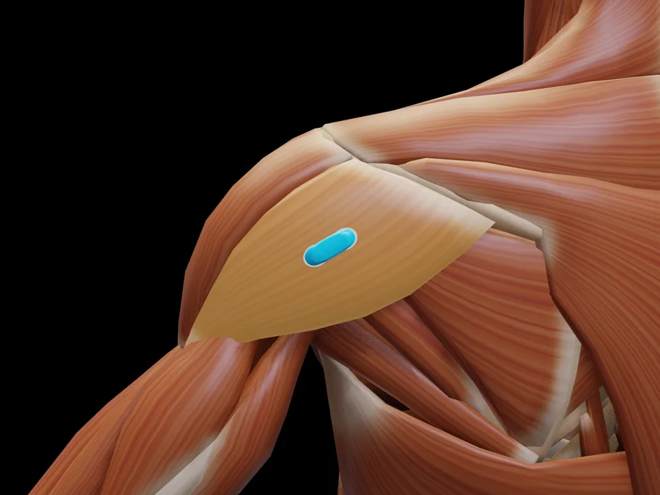
Anatomy & Function
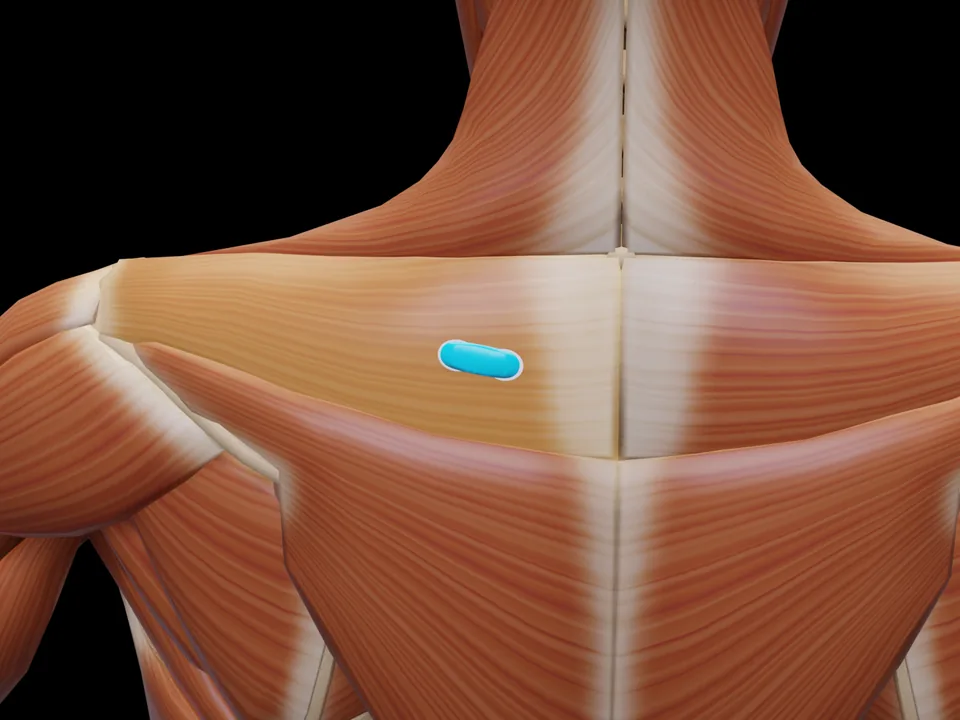
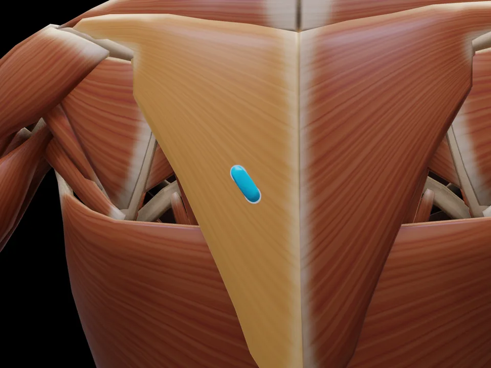
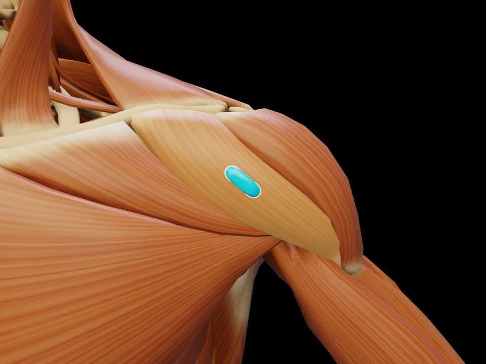
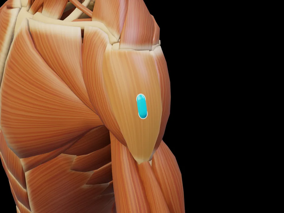
The M. Teres Major (Teres Major) is a muscle of the shoulder region. It is responsible for retroversion, adduction and internal rotation at shoulder joint. This muscle is not covered by the official SENIAM catalogue — placement is based on established clinical literature. This page documents the exact electrode position, anatomical attachments and a standardized test movement based on Hermens et al. (2000).
Innervation after Perotto AO, Anatomical Guide for the Electromyographer, 5th ed. 2011 — an anatomical reference, not an electrode placement recommendation.
Test Movement
Internal rotation of arm against resistance with adducted arm
This muscle has no SENIAM-standardized electrode position. Placement should be based on anatomical knowledge.
MVC reference test (Konrad 2005)
- Position
- Simulated pull-up: elbows at about 90° flexion, check a frontal and a lateral arm position.
- Fixation
- Fixed bar or machine; trunk stabilised.
- Resistance
- Pulling against the fixed bar.
- Control exercise
- The same set-up may also produce maxima for the biceps and the lower trapezius.
The pull-up simulation produces the highest latissimus innervation; the lower trapezius and teres major share the set-up.
Konrad P. The ABC of EMG. Noraxon; 2005, p. 31. Konrad describes this test at group level; it applies to all muscles of the group. · Why and how to normalise to MVC
Measure M. Teres Major activity live
The electrode is placed — what next? easyEMG visualises muscle activity in real time, with SENIAM-compliant placement guidance, up to 4 channels at once and biofeedback training right afterwards.
In rehabilitation, the Teres Major is typically recorded to track progress and retrain activation.