What the Datasheet of an EMG Sensor Really Tells You
An EMG datasheet lists a handful of numbers — input impedance, common-mode rejection, noise, bandwidth, sampling rate, resolution, gain, range. Each of them describes one thing the amplifier does to the few microvolts arriving from the skin, and each has a threshold below which recordings become unreliable. SENIAM's sensor recommendations and Cram's specification table (Ch. 3) give those thresholds. This article explains what every value means in practice, which numbers are "enough", and where marketing figures mislead.
The signal chain in one paragraph
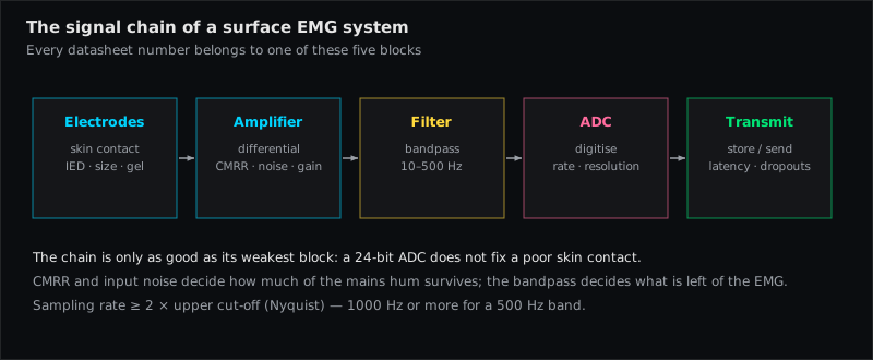
Two electrodes deliver a voltage difference of a few microvolts to a few millivolts. The differential amplifier subtracts the two inputs (cancelling what both share — hum, ECG), multiplies the remainder by the gain, passes it through the bandpass and hands it to an analog-to-digital converter that samples it at the sampling rate with a given resolution. Every specification below describes one link in that chain. Konrad's reminder applies throughout: the amplifier's job is to pick up a signal no bigger than a few millionths of a volt without adding or removing anything — the specifications say how well it does that.
The specifications, one by one
Input impedance
What it is: the resistance the amplifier presents to the electrodes. It must be far higher than the skin impedance, otherwise the amplifier "loads" the electrode interface and attenuates the signal — unequally under the two electrodes, which also degrades common-mode rejection.
Enough: SENIAM asks for > 100 MΩ. Cram's table gives 100 kΩ to 1 GΩ as the range seen in instruments and notes that most commercial devices are at 1 MΩ or better, "more than adequate". Modern amplifiers reach gigaohms; the practical benefit is tolerance of imperfect skin preparation, not a visibly better trace.
Common-mode rejection ratio (CMRR)
What it is: how much of a signal that appears identically at both inputs is suppressed, in decibels. 60 dB = factor 1000, 100 dB = factor 100 000. Mains hum and ECG reach both electrodes almost equally — the CMRR decides how much of them survives.
Enough: SENIAM: > 95 dB. Cram's: 70–180 dB is the range on the market, "the higher the better". Two caveats. First, CMRR is measured under ideal, balanced conditions; unequal skin impedances under the two electrodes convert common-mode noise into a differential signal that no CMRR can remove — which is why skin preparation matters more than the last 20 dB. Second, the figure is frequency-dependent; ask for the value at 50/60 Hz.
Noise floor (input-referred noise)
What it is: the signal the amplifier produces with its inputs shorted — its own electronic noise, in µV RMS over the bandwidth. It is the smallest EMG the system can distinguish from nothing.
Enough: Cram's: 0.1–1.0 µV, most instruments detect 0.5 µV and up. SENIAM: < 1 µV RMS. Compare with Konrad's practical baseline: a well-prepared, relaxed muscle shows 1–3.5 µV mean rectified — so a 1-µV amplifier is at the edge for relaxation work and comfortable for movement work. Read the conditions: "noise" quoted over a narrow band or at a single frequency looks better than the RMS over 10–500 Hz.
Bandwidth (bandpass)
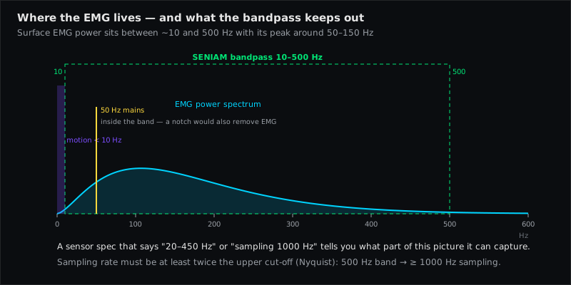
What it is: the frequency range passed, defined by a high-pass and a low-pass corner. Surface EMG power lies mostly between 20 and 250 Hz; Cram's: 20–300 Hz for the general case.
Enough: SENIAM: high-pass 10 Hz (20 Hz for dynamic tasks), low-pass ~500 Hz. Cram's Table 3-2 gives use-dependent choices — 20–1000 Hz general, 20–300 Hz musculoskeletal work, 100–200 Hz relaxation training (which "all but eliminates" ECG at the cost of real signal), 20–600 Hz facial muscles. Watch for fixed, non-adjustable filters and for a high-pass above 20 Hz sold as "noise reduction": it removes fatigue-related low-frequency content along with the artifacts.
Sampling rate
What it is: samples per second at the A/D converter. The Nyquist criterion demands at least twice the highest frequency in the signal.
Enough: SENIAM: ≥ 1000 Hz for a 500-Hz low-pass; 2000 Hz is common and gives headroom. Below 1000 Hz, content above half the sampling rate folds back as aliasing and cannot be removed afterwards. Note the difference between the sensor's internal sampling rate and the rate at which data are transmitted or displayed — a wireless sensor may sample at 2 kHz and stream an envelope at 100 Hz, which is fine for biofeedback but not for spectral analysis.
Resolution (bit depth)
What it is: the number of levels the A/D converter distinguishes. 12 bit = 4096, 16 bit = 65 536, 24 bit = 16.8 million.
Enough: 16 bit is standard; 24 bit removes the need to choose a gain range. What matters is the smallest voltage step: input range divided by the number of levels. A 16-bit converter over ±5 mV resolves 0.15 µV — well below the noise floor, so more bits do not create more information; they only avoid clipping and range switching.
Gain and range
What it is: gain is the amplification factor (Konrad: typically 500–2000); range is the largest amplitude the system can represent before it clips. They are two views of the same setting.
Enough: Cram's Table 3-2: 0–1000 µV RMS for dynamic work, 0–100 µV RMS for relaxation work, and preferably selectable. A system that clips at 500 µV loses every strong contraction of the Gastrocnemius (Medial Head) or Biceps Brachii; a system with only a 5-mV range wastes resolution on a resting Upper Trapezius. Auto-ranging or 24-bit converters make this a non-issue.
Electrode interface and cabling
What it is: the part the datasheet says least about — pre-gelled Ag/AgCl electrodes with ≤ 10 mm conductive diameter, 20 mm inter-electrode distance (SENIAM), and how the leads reach the amplifier. Cram's lists the lead-related artifacts: 50/60-Hz pickup (antenna effect), movement artifact, radio-frequency interference.
Enough: active electrodes or a preamplifier close to the skin, short leads, a fixed 20-mm geometry. Wireless sensors with integrated amplifiers remove the antenna problem entirely — their limiting spec then becomes the transmission rate.
A checklist to compare systems
Cram's suggests keeping one sheet per instrument (Exhibit 3-1). A condensed version:
| Specification | Ask for | Adequate (SENIAM / Cram's) |
|---|---|---|
| Input impedance | value in MΩ or GΩ | > 100 MΩ |
| CMRR | dB at 50/60 Hz | > 95 dB |
| Input-referred noise | µV RMS over 10–500 Hz | < 1 µV RMS |
| Bandwidth | high-pass and low-pass, adjustable? | 10 (20)–500 Hz |
| Sampling rate | Hz at the sensor, and transmitted rate | ≥ 1000 Hz, better 2000 |
| Resolution | bits and input range → µV per step | 16 bit or more |
| Range / gain | max. amplitude before clipping, selectable? | up to 1000 µV RMS dynamic |
| Electrodes | type, diameter, inter-electrode distance | Ag/AgCl, ≤ 10 mm, 20 mm |
| Raw signal access | can you see and export the raw trace? | yes — the signal check needs it |
| Filters applied by software | which, adjustable, documented? | documented and switchable |
The last two rows are not electrical specifications but decide whether you can do the five-step signal check at all. A system that only shows a smoothed envelope hides every artifact from you.
What the numbers do not tell you
- They assume good skin contact. Unequal impedances degrade CMRR in practice regardless of the datasheet — see the ten factors.
- They say nothing about placement. A perfect amplifier over the innervation zone still gives an unstable amplitude.
- Software processing is often undocumented. Envelope window, notch filter, ECG suppression, normalisation defaults — ask, and switch them off when you need the raw signal.
- Marketing terms are not specifications. "Medical grade", "research grade" and "clinical precision" carry no number. Ask for the table above.
Muscles referenced in this article
From placement to measurement
EMG Guide shows where the electrodes go. easyEMG with PicoBlue sensors visualises the signal live afterwards — SENIAM-conform, in real time, with a signal check on screen.
Frequently asked questions
Is a higher CMRR always better?
On paper yes; in practice, beyond about 100 dB the limiting factor is the mismatch of the two skin impedances, not the amplifier. Prepare the skin well and any modern amplifier's CMRR is sufficient.
Do I need 24-bit resolution?
No. 16 bit resolves steps far below the noise floor. 24 bit is convenient because it removes gain-range switching, not because it records "more" EMG.
Which bandwidth should I set?
For movement, strength and fatigue work: 20–500 Hz (10 Hz high-pass if you have very clean, static conditions). For relaxation biofeedback on the trunk and shoulders Cram's suggests 100–200 Hz to suppress ECG — accept that you lose part of the signal. Whatever you choose, keep it constant within a comparison and report it.
Can I trust the noise figure on the datasheet?
Only if it states the bandwidth and the measurement condition (inputs shorted, RMS). Test it yourself: shorted inputs or an electrode pair on a bony, relaxed site should give a flat trace of about 1–2 µV RMS.
Sources
- Criswell E. Cram's Introduction to Surface Electromyography. 2nd ed. 2011 — Ch. 3 "Instrumentation", incl. "How to Check Specifications of SEMG Instruments" and Table 3-2 (pp. 35–62, esp. 57–59).
- Hermens HJ et al. SENIAM — European Recommendations for Surface Electromyography, sensor and signal-processing recommendations. 1999/2000.
- Konrad P. The ABC of EMG. Noraxon; 2005 — pp. 12 (amplification), 21–23 (signal check), 28 (digital filtering).
- Merletti R, Parker PA (eds.). Electromyography — Physiology, Engineering, and Noninvasive Applications. IEEE/Wiley; 2004 — Ch. 5.
- De Luca CJ. The use of surface electromyography in biomechanics. J Appl Biomech. 1997;13:135–163.
Read next
EMG Electrode Placement — The Practical Guide
How to place surface EMG electrodes correctly — SENIAM rules, skin prep, 20 mm spacing, fibre direction, reference electrode, signal check and common mistakes.
10 Factors That Distort Your EMG Signal
Skin, fat, temperature, spacing, innervation zone, crosstalk, ECG, motion, cables, fatigue — ten influences that change an EMG amplitude, and how to control each.
Surface EMG Glossary
54 terms of surface electromyography explained in one sentence each — from amplitude cancellation to volume conduction. With sources (SENIAM, Cram's, Konrad).
All articles →