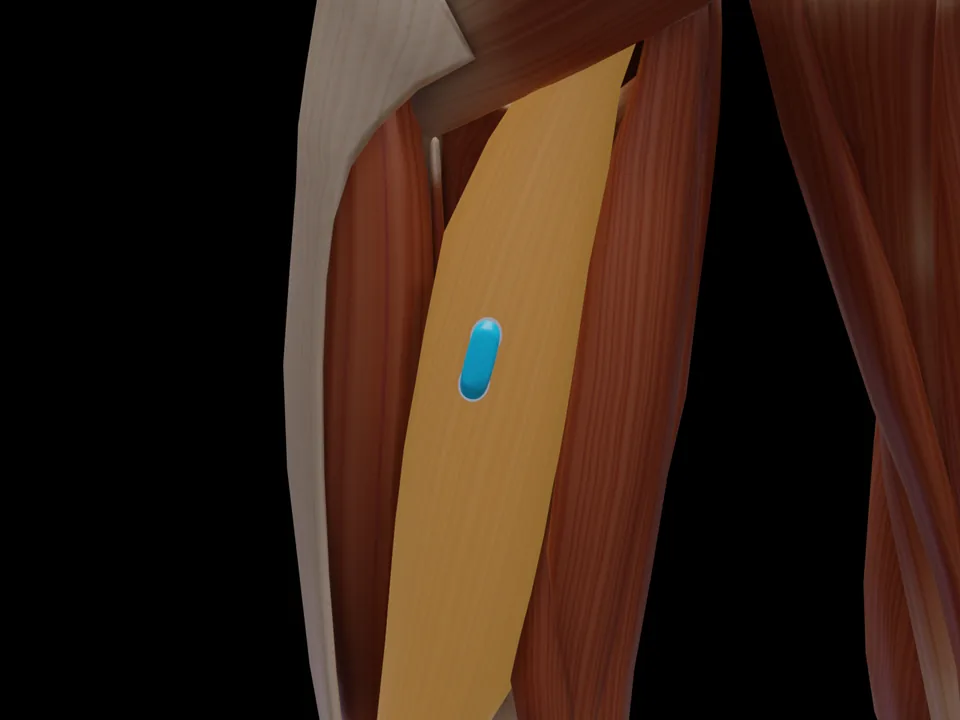
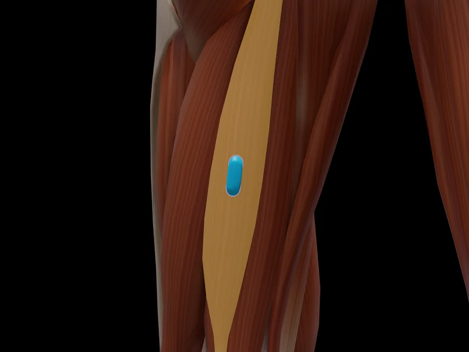
Anatomy & Function
The M. Semimembranosus (Semimembranosus) is a muscle of the thigh posterior region. It is responsible for knee flexion, hip extension, internal rotation of the lower leg with flexed knee. This muscle is not covered by the official SENIAM catalogue — placement is based on established clinical literature. This page documents the exact electrode position, anatomical attachments and a standardized test movement based on Hermens et al. (2000).
Innervation after Perotto AO, Anatomical Guide for the Electromyographer, 5th ed. 2011 — an anatomical reference, not an electrode placement recommendation.
Test Movement
Knee flexion in prone position against resistance with internal rotation
This muscle has no SENIAM-standardized electrode position. Placement should be based on anatomical knowledge.
MVC reference test (Konrad 2005)
- Position
- Prone, unilateral knee flexion held at about 20–30° knee flexion.
- Fixation
- Very good hip fixation (belt or a heavy person holding the pelvis).
- Resistance
- Manual resistance or strap at the heel, knee flexion against it.
- Control exercise
- The prone erector spinae test is the important check exercise.
Konrad tests the hamstrings as one group ("Mm. ischiocrurales"); surface EMG does not separate the medial and lateral heads well.
Konrad P. The ABC of EMG. Noraxon; 2005, p. 32. Konrad describes this test at group level; it applies to all muscles of the group. · Why and how to normalise to MVC
Measure M. Semimembranosus activity live
The electrode is placed — what next? easyEMG visualises muscle activity in real time, with SENIAM-compliant placement guidance, up to 4 channels at once and biofeedback training right afterwards.
In training, what matters for this muscle is targeted activation and left/right symmetry.