Electrode Placement (SENIAM)
50% on line from lateral epicondyle to the ulna
Orientation: Parallel to the forearm (dorsal)
Source:
Farina et al. 2015 ↗
Farina D, Kallenberg LA, Holobar A. Extracting extensor digitorum communis activation patterns using high-density surface electromyography. Front Physiol. 2015;6:279.
Methodological background of the source
Farina et al. (2015) analysed the extensor digitorum using high-density multi-channel EMG (HD-sEMG). Such arrays reveal where on the muscle stable, low-artefact signals can be recorded — the bipolar position used here for the forearm extensor is derived from that.
Recording notes per Criswell 2011, site „Extensor Digitorum“, p. 327
- Placement type
- Quasi-specific site Neighbouring muscles contribute noticeably to the signal — allow for crosstalk.
- Signal check
- Extend the fingers.
- Crosstalk from
- extensor digiti minimi · extensor carpi ulnaris (a distal placement separates the finger extensors better from the wrist flexors)
Based on SENIAM recommendations (Hermens et al. 2000). Not an official SENIAM product. See all references →
· How electrodes are placed (guide)
· Glossary
All 48 muscle protocols as a PDF — free
Print-ready for the lab or clinic: 84 pages of protocols (SENIAM + peer-reviewed), plus the SENIAM table, a placement checklist and a sensor comparison sheet as A4 sheets.
Anatomy
Origin
Lateral epicondyle of the humerus
Insertion
Dorsal aponeurosis of fingers II-V
Innervation
Posterior interosseous nerve from the radial nerve, C7–C8
Test Movement
Finger extension against resistance with extended wrist
This muscle has no SENIAM-standardized electrode position. Placement should be based on anatomical knowledge.
MVC reference test (Konrad 2005)
- Position
- Seated or kneeling in front of a bench with the forearm resting on a stable support.
- Fixation
- Forearm fully supported; wrist free to work against the resistance.
- Resistance
- Manual resistance, a barbell or a cable/belt against the wrist or hand.
- Control exercise
- Use the latissimus dorsi and pectoralis major tests as control exercises.
Konrad treats the forearm flexors and extensors as one group; wrist flexion for the flexors, wrist extension for the extensors.
Konrad P. The ABC of EMG. Noraxon; 2005, p. 31. Konrad describes this test at group level; it applies to all muscles of the group. · Why and how to normalise to MVC
Measure M. Extensor Digitorum activity live
The electrode is placed — what next? easyEMG visualises muscle activity in real time, with SENIAM-compliant placement guidance, up to 4 channels at once and biofeedback training right afterwards.
In therapy, this muscle is usually about tone regulation and consciously relaxing below a threshold.
Frequently Asked Questions
Where do I place the EMG electrode on the M. Extensor Digitorum?
50% on line from lateral epicondyle to the ulna. Orientation: Parallel to the forearm (dorsal). Not covered by SENIAM — placement per Farina et al. 2015.
Which test movement activates the M. Extensor Digitorum?
Finger extension against resistance with extended wrist. It serves as a signal check before recording: with correct electrode placement the EMG amplitude rises clearly during this movement.
Is the M. Extensor Digitorum a SENIAM-recommended muscle?
No. The M. Extensor Digitorum is not included in the official SENIAM catalogue. The electrode position documented here follows the established protocol per Farina et al. 2015.
Related Muscles in This Region
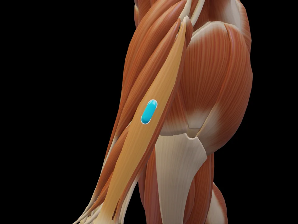 Preview — open the 3D viewer to rotate the model and check neighbouring muscles.
View in 3D Viewer
Preview — open the 3D viewer to rotate the model and check neighbouring muscles.
View in 3D Viewer